Table of Contents
Introduction to CLSM
Confocal Laser Scanning Microscopy (CLSM) is an advanced technique that allows detailed imaging of biological specimens by focusing on thin layers within the sample. This method overcomes the limitations of traditional microscopy techniques, providing high-resolution, sharp images.
Working Principle of CLSM
Confocal microscopy achieves sharp imaging by rejecting out-of-focus light through a pinhole aperture, which collects light only from a thin section of the sample.
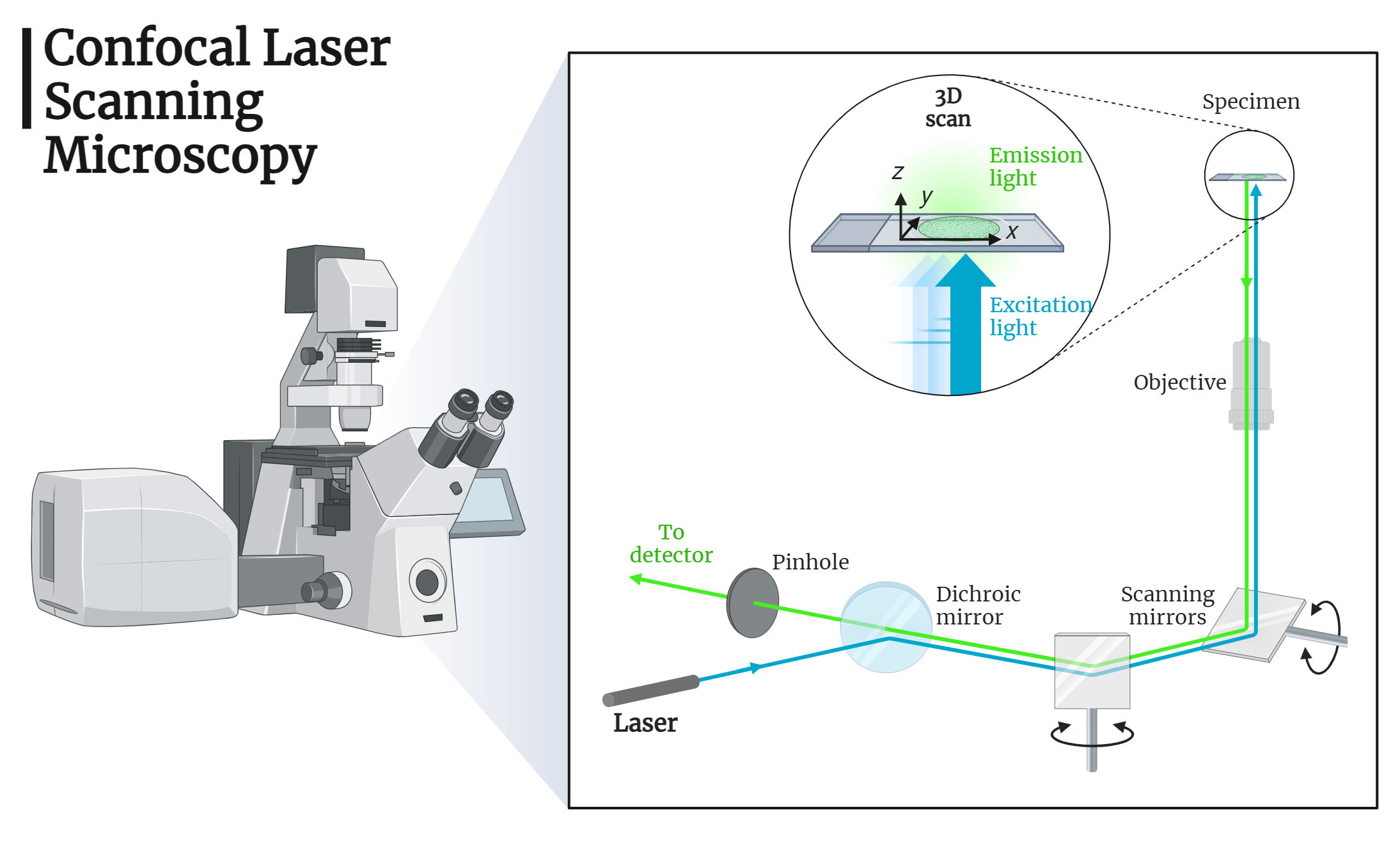
Components and Functions of CLSM
Light Source and Illumination
- Lasers are used as the light sources in confocal microscopes, operating in epi-illumination mode.
- A diverging lens spreads the laser beam to fill the back aperture of the objective lens, which also functions as the condenser.
- The expanded laser light is reflected by a dichroic mirror on the objective, focusing as an intense diffraction-limited spot on the sample.
- The fluorescence from the illuminated spot is collected by the objective and directed through a pinhole aperture to the eyepiece, camera, or detector.
Pinhole Aperture
- The fluorescence light emitted by the sample is focused at the pinhole’s confocal point.
- Light from areas above or below the focal plane is blocked by the pinhole plate, ensuring only in-focus light is detected.
Raster Scanning
- The focused laser spot scans the sample in a raster pattern, collecting light from the entire focal plane.
- The position of the illuminating spot changes, and the pinhole moves to remain confocal with the illuminated spot within the same focal plane.
Optical Sectioning and 3D Reconstruction
- A confocal microscope records the intensity of diffraction-limited spots in a focal plane, providing an optical section of the sample.
- This is visualized as a plot of intensity in two coordinates.
- By obtaining such plots for closely spaced focal planes, three-dimensional reconstruction of the sample is achieved through image stacking.
Emission Filter
- The light passing through the pinhole is filtered by an emission filter before reaching the detector, ensuring only the desired wavelengths are detected.
Conclusion
Confocal Laser Scanning Microscopy (CLSM) is a powerful tool for high-resolution imaging of biological specimens. By utilizing lasers, pinhole apertures, and raster scanning, CLSM provides sharp, detailed images and enables three-dimensional reconstruction of samples. This technique offers significant advantages for researchers, allowing for precise localization and visualization of cellular structures and molecules. Understanding the principles and components of CLSM paves the way for advancements in biological imaging and scientific research.
