Table of Contents
Introduction to electron microscopy
In 1924, Louis de Broglie proposed that particles exhibit wave like properties. This theory was experimentally confirmed in 1927 by Davisson and Germer, as well as Thomson and Reid, through electron diffraction experiments. This paved the way for the development of electron microscopy, with Knoll and Ruska publishing the first images using electrons within five years.
de Broglie Equation
The wavelength (λ) of a particle with velocity (v) and momentum (p) is given by the de Broglie equation:

where h is the Planck’s constant, m is the mass of the particle, and v is the particle’s velocity.
Electron Acceleration in Microscopy
In an electron microscope, electrons are accelerated by a potential difference (V). The kinetic energy of the accelerated electrons is given by:

where e is the charge of the electron, m0 is its rest mass, and v is its velocity. Substituting for v in the de Broglie equation, we get:

Relativistic Effects
At very high accelerating potentials (greater than ~100 kV), the electron’s velocity approaches the speed of light (c), necessitating the incorporation of relativistic effects. The modified equation for wavelength is:

Substituting the constants into this equation gives a more practical wavelength calculation.
Resolution in Electron Microscopy
Unlike light microscopy, electron microscopy requires a very high vacuum (pressure ~10−5 Pa or less) to prevent electron scattering by air molecules. The mean free path in an electron microscope needs to be ~1-2 meters, necessitating this high vacuum. Magnetic fields act as lenses in focusing electron beams. Under high vacuum conditions, the refractive index n≈1.
The equation for resolution (R) is simplified to:

where α is the aperture angle. Assuming α=5∘ (0.1 radian) and operating at a voltage of 100 kV, the theoretical resolution is around 2.26 pm. In practice, resolutions better than 0.2 nm are rare due to lens aberrations.
Electron Sources and Lenses
Two main methods for generating electrons in electron guns are thermionic emission and field emission. Most electron microscopes use thermionic emission from a heated filament, with tungsten being the most common filament material due to its cost-effectiveness and efficiency. Lanthanum hexaboride (LaB6) can be used for higher brightness, achieving ~1010 A·m−2·sr−1. Field emission guns, which use a single crystal tungsten filament with a fine tip, provide even higher brightness (> 1013 A·m−2·sr−1) by applying a strong electric field.
Magnetic lenses, the core of an electron microscope, focus the electron beam. The Lorentz force law describes the deflection experienced by electrons in a magnetic field:

Scattering of Electrons
Electron microscopy relies on interactions between electrons and matter, resulting in changes in the electrons or the emission of new electrons with different energies. These interactions include:
Elastic Scattering In elastic scattering, electrons change their trajectories without losing energy. This process results in a strong forward peak, especially in thin specimens.
Inelastic Scattering Inelastic scattering involves electrons losing energy as they interact with the specimen. This energy loss results in various inelastic scattering processes.
Secondary Effects Primary electrons can cause several secondary effects:
- Secondary Electrons: Low energy electrons (below 50 eV) ejected from atoms in the specimen. These electrons, produced abundantly, are crucial for scanning electron microscopy (SEM).
- Backscattered Electrons: Primary electrons that retain significant energy and are scattered back from the specimen surface. The intensity of backscattered electrons is higher in samples with larger atomic numbers.
- Cathodoluminescence: Emission of light due to electron-induced excitation, where an electron knocks off a valence electron, creating an electron-hole pair. When an electron falls back into the hole, it releases energy as light.
- X Rays : Generated when an electron from a higher energy shell fills a vacancy in a lower energy shell, releasing energy in the X-ray wavelength region.
Types of Electron Microscopes
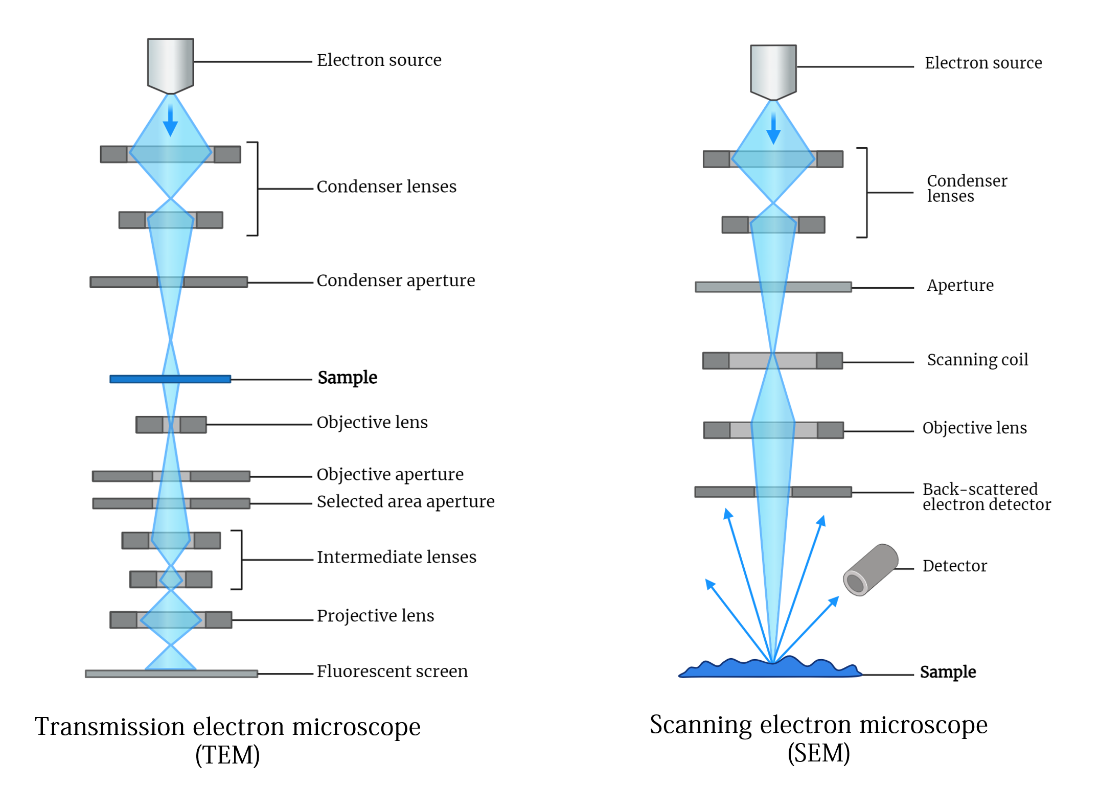
- Scanning Electron Microscopes (SEM): Detect electrons scattered from the surface of a specimen.
- Transmission Electron Microscopes (TEM): Detect electrons transmitted through a specimen.
Conclusion
Electron microscopy has revolutionized our ability to observe and understand the microscopic world. Building on the foundational theories of de Broglie and the pioneering experiments of Davisson, Germer, Thomson, and Reid, it enables scientists to achieve remarkable resolutions and insights into the structure and behavior of materials at the atomic level. The principles of electron acceleration, the role of magnetic lenses, and the varied scattering processes create a robust framework for detailed imaging. Advanced electron sources further enhance the capabilities of electron microscopes. This sophisticated technology continues to be an invaluable tool in numerous fields, driving forward scientific research and discovery.
