Table of Contents
Introduction to Fluorescence Microscope:
A fluorescence microscope is a type of microscope that uses a fluorescence technique to produce high-resolution images of a sample. It is an essential tool for many fields of science and technology, including biology, chemistry, and materials science.
Definition of Fluorescence Microscope:
Fluorescence microscopy is a type of microscopy in which the sample is excited by a specific wavelength of light and the emitted light is detected and captured by a camera.
Discovery:
- This microscope was first developed in the early 20th century by August Köhler.
- Köhler was awarded the Nobel Prize in Physics in 1931 for his work in developing the fluorescence microscope.
Principal:
- This microscope works by using a specific wavelength of light to excite fluorescence in a sample.
- The fluorescence emitted by the sample is then collected by the microscope and used to create an image of the sample.
- This allows for high-resolution images to be produced.
Components:
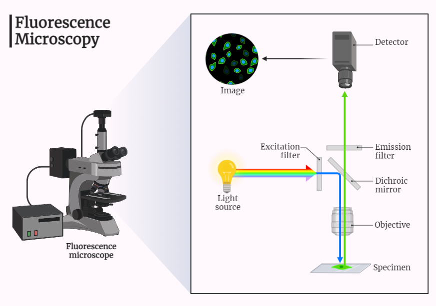
- The main components of a fluorescence microscopy include the light source, the objective lens, the filter, the detector, and the computer.
- The light source is used to excite fluorescence in the sample.
- The objective lens focuses the light on the sample.
- The filter is used to filter out the excitation light and only allow the fluorescence to pass through.
- The detector is used to detect the fluorescence emitted by the sample.
- The computer is used to control the microscope and to process the images.
Steps:
- The first step in using a fluorescence microscope is to prepare the sample. This can involve staining the sample with a fluorescent dye or protein.
- Next, the sample is placed on the microscope stage and the light source is turned on.
- The light source is directed at the sample and the detector measures the fluorescence emitted by the sample.
- The filter is used to filter out the excitation light and only allow the fluorescence to pass through.
- The fluorescence is then collected by the microscope and used to create an image of the sample.
Applications:
- These microscopes are used in a wide variety of fields, including biology, chemistry, and materials science.
- They are used to study the structure and composition of cells and other biological samples, to analyze chemical compounds, and to study the properties of materials.
- They are also used in medical research to study the structure of human tissue and in biotechnology to study the structure of cells and proteins.
Conclusion:
The fluorescence microscope is an essential tool for many fields of science and technology. Its ability to produce high-resolution images of a sample has led to many important discoveries and advancements in a wide variety of fields. This microscope allows to detect specific molecules or structures within the samples, and because of its selectivity, it is particularly useful in studying biological samples and their components in details.
