Table of Contents
Introduction to fluorescence microscopy
- Fluorescence microscopy has significantly evolved since its early applications in the 20th century.
- This technique directly detects fluorescence in the visible spectrum of electromagnetic radiation, unlike other forms of light microscopy that require special optics to enhance contrast.
- Most biomolecules do not naturally fluoresce in the visible region, so extrinsic fluorescent probes are used for studying cellular features with high specificity.
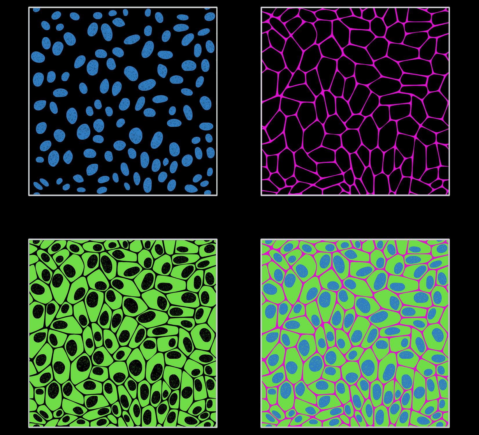
Immunofluorescence
- Immunofluorescence utilizes the high specificity of antibodies to target cellular markers and organelles.
- For cell surface markers, cells are treated with fluorescently labeled antibodies and examined under a microscope.
- For intracellular targets, cells are usually fixed and permeabilized to allow antibody penetration, providing detailed distribution information of the target molecules within the cells.
- This technique is limited to non-living cells due to the need for fixation and permeabilization.
Alternative Approaches
- Small fluorescent dyes can translocate across biological membranes and bind to cellular targets with high specificity.
- Directly labeling molecules with fluorescent tags, such as carboxyfluorescein linked to synthetic peptides, allows for microscopic studies.
- However, direct labeling is not always suitable for specific intracellular molecules.
GFP and Variants
- The discovery of green fluorescent protein (GFP) and its variants has revolutionized protein labeling inside cells.
- GFP can be tagged to either end of a protein, which is crucial for proteins with N-terminal localization signals to maintain proper localization.
Fluorescence Microscope
- An epifluorescence microscope uses a single lens to illuminate the specimen and collect fluorescence light, made possible by the incorporation of a dichroic mirror.
- The microscope features a high-power lamp source, such as a mercury or xenon arc lamp, which provides excitation radiation.
- The dichroic mirror reflects excitation radiation and transmits emitted fluorescence to the ocular lens.

Upright vs. Inverted Microscopes
- In upright microscopes, the objective turret is fixed, and the sample stage moves to focus the image.
- In inverted microscopes, the sample stage is fixed while the objective turret moves, offering better sample manipulation, mechanical stability, and suitability for attaching TIRF accessories.
Autofluorescence
- Many essential cellular molecules, such as B-vitamins, flavins, cytochromes, and nucleotides (FMN, FAD, NADH), are naturally fluorescent.
- Autofluorescence from these molecules is most intense when cells are excited in the UV/blue region and can sometimes be mistaken for specific signal fluorescence.
TIRF
- Total internal reflection fluorescence (TIRF) selectively excites molecules near the slide or culture plate surface, producing fluorescence from a thin sample layer.
- This technique is particularly useful for studying membrane proteins.
Conclusion
- Fluorescence microscopy, including techniques like immunofluorescence and TIRF, is essential in biological research.
- By utilizing fluorescent probes, antibodies, and proteins like GFP, researchers can gain detailed insights into cellular structures and functions.
- Understanding these techniques paves the way for further advancements in scientific research and our comprehension of cellular processes.
