Table of Contents
Introduction to Gel Electrophoresis:
Gel electrophoresis is a widely used technique in molecular biology for the separation and analysis of nucleic acids and proteins. It is based on the principle of utilizing an electric field to move charged molecules through a gel matrix, allowing for their separation based on size, charge, and shape. Gel electrophoresis plays a crucial role in various applications, including DNA sequencing, genetic fingerprinting, and protein analysis.
Principles of Gel Electrophoresis:
The key principles of gel electrophoresis are as follows:
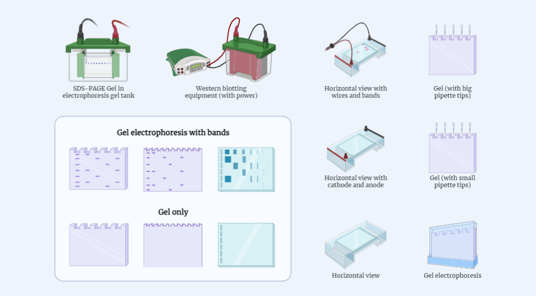
- Electric Field: An electric field is applied across a gel matrix, typically made of agarose or polyacrylamide, which creates a migration pathway for charged molecules.
- Gel Matrix: The gel matrix serves as a molecular sieve, slowing down the movement of molecules based on their size and shape. Agarose gels are commonly used for separating larger DNA fragments, while polyacrylamide gels are suitable for smaller DNA fragments and proteins.
- Migration: Charged molecules migrate through the gel matrix in response to the electric field. Negatively charged molecules move towards the positive electrode (anode), while positively charged molecules move towards the negative electrode (cathode).
- Separation: The gel matrix allows for the separation of molecules based on their size. Smaller molecules can move more easily through the gel matrix, resulting in faster migration and appearing closer to the positive electrode, while larger molecules migrate slower and appear closer to the starting point.
Procedure of Gel Electrophoresis:
The general steps involved in gel electrophoresis are as follows:
- Gel Preparation: Prepare the gel matrix by melting and solidifying agarose or polyacrylamide gel, creating a gel slab or a gel column.
- Sample Preparation: Mix the nucleic acid or protein sample with a loading buffer that contains tracking dyes to visualize the migration of molecules during electrophoresis.
- Loading: Load the sample into wells or slots made on the gel using a pipette. Include appropriate controls for comparison.
- Electrophoresis: Submerge the gel in a buffer solution that conducts electricity and connect the power supply. Apply a voltage across the gel, allowing molecules to migrate through the gel matrix based on their charge and size.
- Visualization: After electrophoresis, stain the gel with dyes or use specific detection methods to visualize the separated molecules. DNA fragments are commonly visualized using DNA-intercalating dyes, while proteins may require specific staining or immunodetection techniques.
Applications:
Gel electrophoresis finds extensive applications in various fields, including:
- DNA Analysis: It is essential for DNA sequencing, genotyping, and genetic fingerprinting. It enables the separation and identification of DNA fragments of different sizes, aiding in DNA analysis.
- Protein Analysis: This technique is used to analyze and characterize proteins based on their size, charge, and molecular weight. It plays a crucial role in protein purification, studying protein-protein interactions, and assessing protein expression levels.
- RNA Analysis: Gel electrophoresis is utilized for RNA analysis, such as determining the integrity and size of RNA molecules and analyzing gene expression through RNA profiling.
- Clinical Diagnostics: It is employed in clinical settings to detect genetic disorders, such as sickle cell anemia or thalassemia, by analyzing DNA or RNA fragments associated with specific mutations.
Conclusion:
Gel electrophoresis is a fundamental technique in molecular biology, enabling the separation and analysis of nucleic acids and proteins based on their size and charge. It has revolutionized various fields, from DNA sequencing to protein analysis, and continues to be a cornerstone in scientific research and clinical diagnostics.