Table of Contents
Introduction:
The kidney is a very vital organ responsible for maintaining the body’s fluid and electrolyte balance, regulating blood pressure, and filtering waste products from the blood. Understanding the anatomy of the kidneys is essential for comprehending their functions and the diseases that can affect them. This study note will provide an in-depth explanation of the anatomy of the human kidneys.
Structure of the Kidney:
- The kidneys are bean-shaped organs located in the upper abdominal cavity, on either side of the spine. They are retroperitoneal, meaning they lie behind the peritoneum. Each kidney is approximately 11 cm in length and is enclosed by a fibrous capsule that provides protection.
- External Anatomy: The external surface of the kidney is convex, while the inner surface is concave, forming the renal hilum. The renal hilum is the entry and exit point for blood vessels, nerves, and the ureter, which carries urine from the kidney to the bladder. The renal hilum also serves as the site for lymphatic drainage.
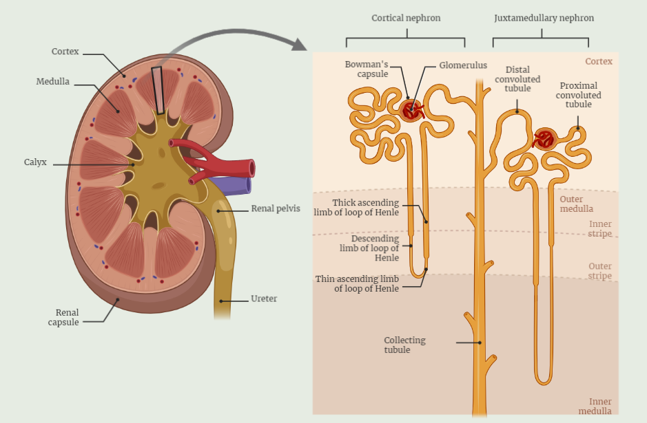
- Internal Anatomy: Internally, each kidney is composed of two distinct regions: the renal cortex and the renal medulla.
- Renal Cortex: The renal cortex is the outer region of the kidney and contains numerous renal corpuscles, which are responsible for the initial filtration of blood. The renal cortex also houses the proximal and distal convoluted tubules, which are involved in the reabsorption and secretion processes.
- Renal Medulla: The renal medulla is the inner region of the kidney and consists of renal pyramids. These pyramids contain tubules that transport urine to the renal pelvis. The renal medulla plays a crucial role in maintaining the concentration and dilution of urine.
Nephron:
The nephron is the functional unit of the kidney and is responsible for urine formation. Each kidney contains millions of nephrons. A nephron consists of a renal corpuscle (composed of the glomerulus and Bowman’s capsule) and a renal tubule (proximal convoluted tubule, loop of Henle, and distal convoluted tubule). The nephron is responsible for filtration, reabsorption, and secretion processes that occur within the kidneys.
Blood Supply:
The kidneys receive a significant blood supply to facilitate their functions. Renal arteries, arising from the abdominal aorta, bring oxygenated blood to the kidneys. Within the kidneys, the renal arteries branch into smaller arteries, eventually forming a network of capillaries known as the glomerulus. After filtration, blood is carried away from the kidneys by the renal veins, which ultimately drain into the inferior vena cava.
Conclusion:
Understanding the anatomy of the kidneys is essential for comprehending their functions and the various processes involved in urine formation. The kidneys’ external and internal structures, including the renal cortex, renal medulla, and nephrons, play crucial roles in maintaining fluid and electrolyte balance. The kidneys’ intricate blood supply ensures the filtration and removal of waste products from the blood. Knowledge of kidney anatomy provides a foundation for studying renal physiology and diagnosing and treating kidney-related disorders.
