Table of Contents
Introduction to Magnetic Resonance Imaging (MRI)
Magnetic Resonance Imaging (MRI) is a non-invasive medical imaging technique that uses magnetic fields and radio waves to create detailed images of the internal structures of the body. MRI is widely used for diagnostic purposes, as it is able to provide high-quality images of soft tissues, organs, and bones. Unlike X-rays, CT scans, and other imaging techniques that use ionizing radiation, MRI does not expose patients to harmful radiation.
Magnetic Resonance Imaging (MRI) Principles and Technology
The basic principle of MRI involves the use of magnetic fields and radio waves to excite the protons in the body’s tissues. When these protons are excited, they emit radio waves of their own, which can be detected by the MRI machine. By measuring the intensity of these radio waves, a computer can create a detailed image of the internal structures of the body.
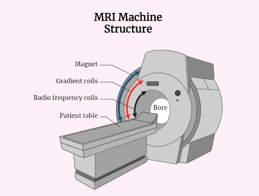
The MRI machine consists of a large magnet, which generates a strong magnetic field. The patient lies on a table that slides into the machine, and a series of radio wave pulses are sent through the body. As these pulses interact with the protons in the tissues, they produce a signal that can be detected by the machine. The computer then uses this signal to create a detailed image of the internal structures of the body.
Physics of MRI :
- MRI works by utilizing the principle of nuclear magnetic resonance (NMR).
- In NMR, the nuclei of certain atoms, such as hydrogen, have a magnetic moment that can be aligned with an external magnetic field.
- When a radio frequency (RF) pulse is applied, the nuclei absorb energy and become excited.
- As the nuclei return to their original state, they emit energy in the form of radio waves, which can be detected and used to create an image.
Magnetic Field:
- The magnetic field used in MRI is generated by superconducting magnets, which are cooled to very low temperatures to maintain their superconductive properties.
- The strength of the magnetic field is measured in tesla (T), and most MRI machines have a field strength of 1.5-3 T.
- A stronger magnetic field results in a higher signal-to-noise ratio, which improves image quality.
RF Pulse:
- The RF pulse is used to excite the nuclei and cause them to emit energy.
- The pulse is delivered by a coil that is placed in close proximity to the area of the body being imaged.
- The pulse is of a specific frequency, and the duration and strength of the pulse can be adjusted to optimize the image.
Gradient Fields:
- Gradient fields are used to selectively excite specific regions of the body and create a spatial encoding of the image.
- Three gradient fields are used: the x, y, and z gradients.
- These gradients are applied during the data acquisition process and affect the position and frequency of the nuclei that are excited.
Sequences:
- Different sequences can be used to optimize the image for different types of tissue and pathology.
- The most common sequences are T1-weighted, T2-weighted, and proton density-weighted.
- Each sequence has its own unique characteristics, such as contrast and resolution, and is used for specific purposes.
Types of MRI
There are several different types of MRI scans that are used for different purposes. Some of the most common types of MRI include:
- T1-weighted MRI: This type of MRI is used to create high-resolution images of the body’s soft tissues. T1-weighted images are useful for detecting tumors, injuries, and other abnormalities in the body.
- T2-weighted MRI: This type of MRI is used to create images that highlight the body’s fluid-filled structures, such as the brain and spinal cord. T2-weighted images are useful for detecting infections, inflammation, and other conditions that affect the body’s fluids.
- Functional MRI (fMRI): This type of MRI is used to measure changes in the brain’s blood flow and oxygenation levels. fMRI is useful for studying brain function and mapping the areas of the brain that are activated during different tasks.
Applications of MRI
MRI is used for a wide range of medical applications, including:
- Diagnosis of diseases and injuries: MRI is often used to diagnose conditions such as cancer, heart disease, and neurological disorders. It can also be used to detect injuries to the muscles, tendons, and ligaments.
- Monitoring the progression of disease: MRI can be used to monitor the progression of diseases such as multiple sclerosis, Alzheimer’s disease, and Parkinson’s disease.
- Planning and guiding surgical procedures: MRI can be used to plan and guide surgical procedures, such as the removal of tumors and the repair of spinal injuries.
- Research and development: MRI is used extensively in medical research, particularly in the study of the brain and neurological disorders.
Advantages and Disadvantages of MRI
Advantages:
- Non-invasive: MRI does not require any incisions or injections, making it a non-invasive imaging technique.
- Safe: MRI does not expose patients to ionizing radiation, which can be harmful.
- High-quality images: MRI is able to create high-quality images of the body’s soft tissues, which can be useful for detecting diseases and injuries.
Disadvantages:
- Costly: MRI machines are expensive to purchase and maintain, which can make them inaccessible to some patients.
- Time-consuming: MRI scans can take up to an hour or more to complete, which can be inconvenient for some patients.
- Claustrophobia: Some patients may feel claustrophobic during an MRI scan, as they are required to lie still in a confined space for an extended period of time.
Conclusion
MRI is a powerful imaging technique that has revolutionized the field of diagnostic medicine. It provides high-quality images of the internal structures of the body, making it an invaluable tool for diagnosing diseases and injuries. Despite its many advantages, MRI does have some drawbacks, including its cost and the time required to complete a scan. Nonetheless, the benefits of MRI far outweigh its disadvantages, and it is likely to remain an essential tool for medical diagnosis and research for many years to come.
