Table of Contents
Introduction
Microscopy refers to the tools and techniques used to visualize microscopic objects and macromolecules. It encompasses a variety of methods for studying biomolecules and biological processes. Light microscopy, the simplest form, utilizes electromagnetic radiation for magnification.
Principles of Microscopy
Geometrical Optics
Light microscopy involves using glass to bend and focus light. Refraction, the bending of light, occurs because light travels at different velocities in different materials. The refractive index measures the light’s velocity in a material, and Snell’s law describes the bending of light as it enters from one material to another.
A convex lens is the simplest form of a microscope. It magnifies an object by bending light rays parallel to its optical axis through the lens’s focus. A compound microscope, which uses two lenses, magnifies the image first with one lens and then with a second lens. The overall magnification is the product of the magnifications of the objective and ocular (eyepiece) lenses:
Mfinal=Mobjective×Mocular
Resolution
Resolution, defined as the minimum distance between point objects that can be distinguished (dmin), is crucial in microscopy. A lower dmin means higher resolution. For light microscopes, the resolution is higher at the blue end of the visible spectrum compared to the red end, assuming other parameters are constant. The theoretical limit for dmin in high refractive index conditions (typically nmax=1.4 for oil) is approximately 0.17 µm, assuming λ=400 nm and sinα=1. This means light microscopes cannot resolve particles closer than ~0.17 µm. Reducing the wavelength of the light source can improve resolution.
Parts of a Light Microscope
A light microscope has several key components. The light source, typically a lamp, is focused on the specimen by the condenser. The objective lens collects light diffracted by the sample to generate a real, magnified image, which is further magnified by the eyepiece.
Types of Light Microscopy
Bright-field Microscopy
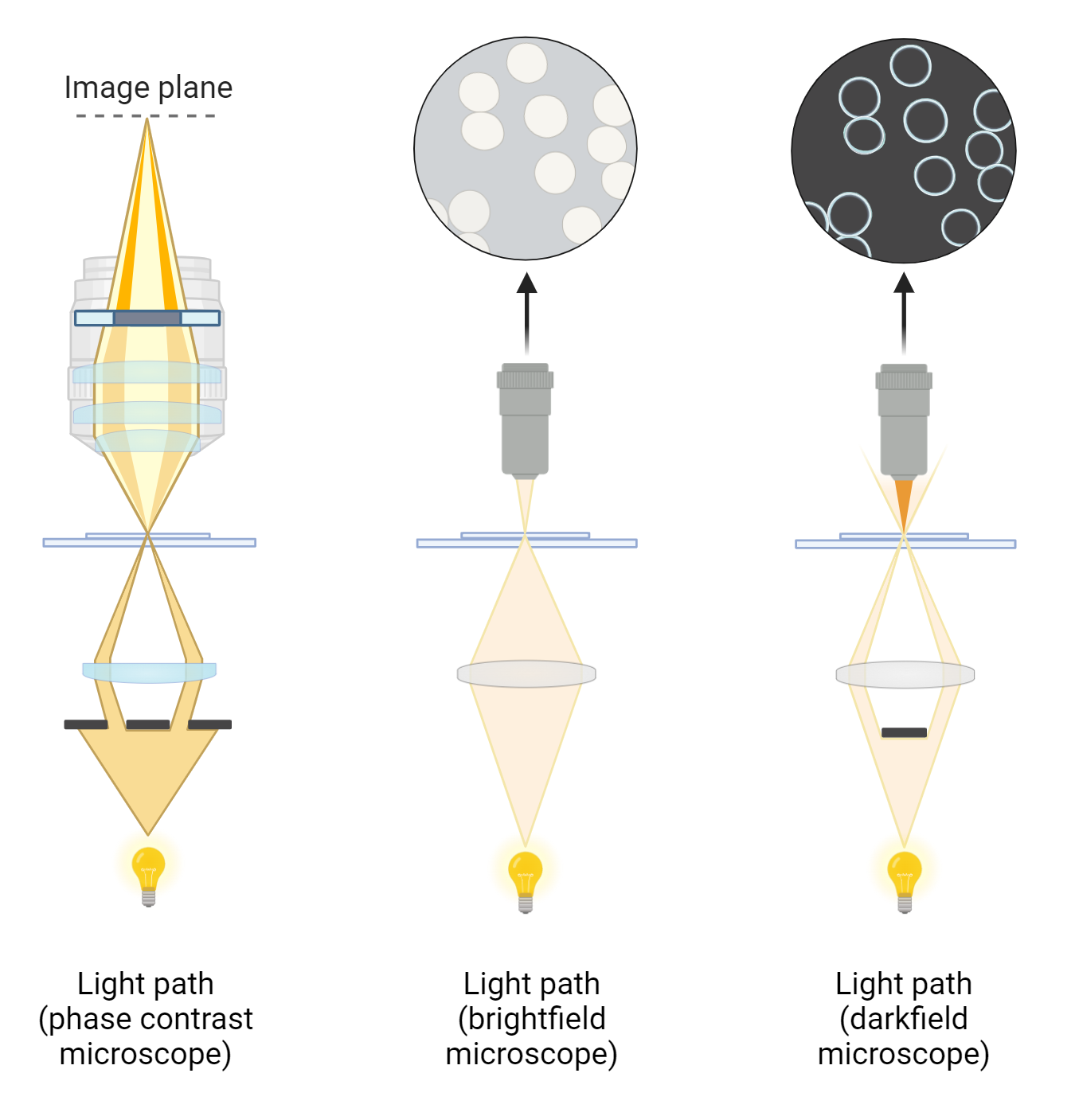
In bright-field microscopy, both diffracted and undiffracted light are collected by the objective lens, producing an image against a bright background. Many biological samples are transparent, resulting in low contrast images. Staining specimens with dyes increases contrast. Intrinsically colored samples, such as erythrocytes, can be observed directly without staining.
Dark-field Microscopy
Dark-field microscopy enhances contrast by eliminating undiffracted light. Light illuminates the specimen from a ring at an oblique angle. Without a specimen, no light is collected by the objective lens. When a specimen is present, it diffracts light, which the objective lens collects, creating a bright image against a dark background.
Phase Contrast Microscopy
Phase contrast microscopy provides higher contrast than bright-field and dark-field methods. It uses both diffracted and undiffracted light to generate images. Light from a ring, called a condenser annulus, illuminates the specimen. The phase plate at the back of the objective lens alters the phase of the undiffracted light by π2 and decreases that of diffracted light by π2, creating a total phase difference of π. The intensity of undiffracted light is reduced to ~30% by a semi-transparent metallic film on the phase plate. This phase difference results in destructive interference, diminishing light intensity. Any phase change caused by the specimen converts into an amplitude signal, increasing contrast
Conclusion
Microscopy, specifically light microscopy, is an essential tool in biological research, allowing for the visualization of microscopic objects and macromolecules. Through principles of geometrical optics, light microscopes use glass lenses to bend and focus light, with different forms of microscopy, such as bright-field, dark-field, and phase contrast, each offering unique ways to enhance image contrast and resolution. The use of techniques like liposomal delivery in cell biology and organismal studies exemplifies the practical applications and advancements in microscopy. Understanding these fundamentals paves the way for more in-depth exploration and innovation in scientific research.

