Table of Contents
Overview
Multiphoton laser scanning microscopy is an advanced imaging technique that leverages the simultaneous absorption of multiple photons by a fluorophore. This process allows the excitation of fluorophores using longer wavelength light (lower energy) than what is typically required.
Key Principles
Two-Photon Absorption In a two-photon absorption process, a fluorophore absorbs two photons simultaneously, each providing half the energy needed for excitation. The wavelength of the absorbed light is twice that of the light used in single-photon excitation.
Challenges and Solutions
- Low Probability of Absorption: The chance of two photons being absorbed simultaneously is very low, which necessitates using very intense light sources.
- Intense Light Sources: Pulsed infrared lasers, such as Titanium:sapphire lasers operating at 800 nm, are used to achieve the required intensity. These lasers can excite fluorophores with a maximum absorption wavelength of around 400 nm.
Advantages of Multiphoton laser scanning Microscopy
- Deeper Tissue Imaging: Biological specimens absorb near-infrared (near-IR) radiation less than ultraviolet (UV) and blue-green radiation, allowing for imaging of thicker samples.
- Clear Separation of Emission and Excitation: Exciting fluorophores at twice their absorption wavelength ensures that emitted radiation is easily distinguishable from excitation radiation and Raman scattering. This clear separation enhances signal detection.
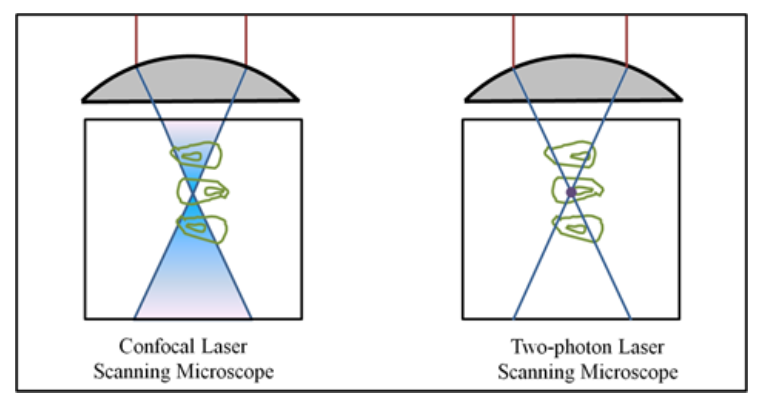
- Focal Plane Excitation: The probability of two-photon absorption depends on the square of the light intensity. High photon density required for excitation is achieved only at the focal point, minimizing excitation outside this plane. Therefore, a multiphoton microscope does not need a pinhole for recording confocal images.
Applications
Multiphoton fluorescence microscopy is ideal for studying living tissues in-depth, making it a powerful tool in biological and medical research.
- Neuroscience: It helps visualize neuronal activity and structures in live brain tissues, aiding the study of brain functions and neural networks.
- Cancer Research: Used to observe tumor growth and behavior at a cellular level, offering insights into cancer progression and treatment effectiveness.
- Developmental Biology: Allows the examination of living embryos and developmental processes over time without damaging the specimens.
- Stem Cell Research: Facilitates the study of stem cell differentiation and their interactions within tissues, which is critical for regenerative medicine.
- Immunology: Used to track immune cells and their responses in real-time, enhancing understanding of immune system functions and diseases.
These applications demonstrate the versatility and power of multiphoton microscopy in advancing our understanding of complex biological systems.
