Table of Contents
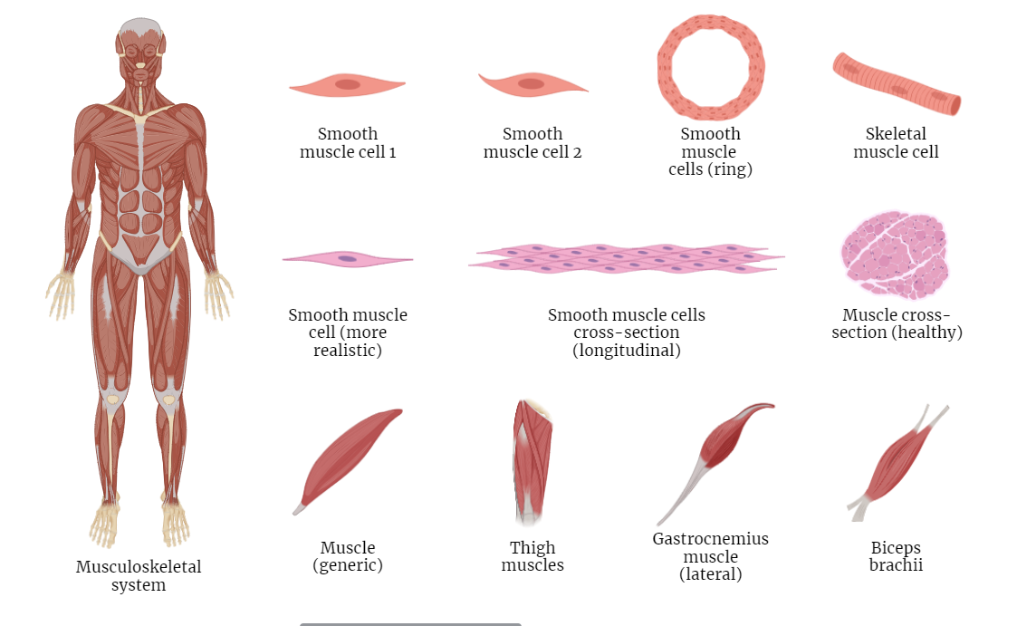
Introduction to skeletal muscle organization
- Definition: Skeletal muscles are composed of muscle fibers that contract to generate movement.
- Hierarchy: Organized from the whole muscle to molecular components.
Gross Anatomy
- Muscle: An organ made up of muscle tissue, blood vessels, tendons, and nerves.
- Fascicles: Bundles of muscle fibers within the muscle.
Muscle Fibers
- Myofibers: Long, cylindrical cells that make up fascicles.
- Sarcolemma: The cell membrane of a muscle fiber.
- Sarcoplasm: The cytoplasm of a muscle fiber.
Myofibrils
- Definition: Rod-like units within muscle fibers.
- Sarcomeres: The basic contractile units of myofibrils.
- Myofilaments: Actin (thin) and myosin (thick) filaments that slide past each other to produce contraction.
Detailed Sarcomere Structure
- Z-disc: Marks the lateral boundary of each sarcomere.
- Function: Anchors the thin filaments and connects adjacent myofibrils.
- I-band: The lighter area containing only thin filaments.
- Function: Changes width during muscle contraction and relaxation.
- A-band: The dark area containing the entire length of thick filaments.
- Overlap Region: Where thick and thin filaments overlap.
- Function: Remains constant in width during contraction.
- H-zone: The central part of the A-band with only thick filaments.
- Function: Disappears during maximum muscle contraction.
- M-line: Located in the center of the H-zone; contains proteins that hold thick filaments in place.
- Function: Maintains the alignment of the sarcomere during contraction.
Molecular Components of Muscle Contraction
- Actin Filaments (Thin Filaments):
- G-actin (Globular Actin): Polymerizes to form F-actin strands.
- F-actin (Filamentous Actin): Two F-actin strands twist together to form the thin filament.
- Tropomyosin: Blocks the myosin-binding sites on actin in a relaxed muscle.
- Troponin Complex: Binds to tropomyosin; moves it away from myosin-binding sites when calcium ions are present.
- Myosin Filaments (Thick Filaments):
- Myosin Heavy Chains: Form the backbone of the filament.
- Myosin Heads: Bind to actin to form cross-bridges; contain ATPase activity for energy transduction.
- Titin: Connects myosin filaments to the Z-disc; provides elasticity and stability.
- Regulatory Proteins:
- Troponin: Binds calcium ions and initiates the contraction process by moving tropomyosin.
- Tropomyosin: Regulates the access of myosin to actin binding sites.
Contraction Mechanism
- Crossbridge Formation: Myosin heads bind to actin forming crossbridges.
- Power Stroke: Myosin heads pivot, pulling actin filaments toward the center of the sarcomere.
- ATP Binding and Myosin Detachment: A new ATP molecule binds to the myosin head, causing it to release from actin.
- ATP Hydrolysis and Cocking of Myosin Head: ATP is hydrolyzed, re-energizing the myosin head and returning it to the cocked position.
These details provide a more comprehensive understanding of the sarcomere’s structure and the molecular components involved in muscle contraction. If you need further clarification or additional information, feel free to ask!
Neuromuscular Junction
- Motor End Plate: The site where the motor neuron communicates with the muscle fiber.
- Acetylcholine (ACh): The neurotransmitter that triggers muscle contraction.
Excitation-Contraction Coupling
- Action Potential: An electrical signal that triggers muscle contraction.
- Calcium Ions: Released from the sarcoplasmic reticulum, initiating the sliding filament mechanism.
Regeneration and Repair
- Satellite Cells: Stem cells that aid in muscle repair and growth.
- Hypertrophy: Increase in muscle size due to an increase in myofibril size.
Clinical Correlation
- Muscular Dystrophy: A group of diseases that cause progressive weakness and loss of muscle mass.
- Myopathies: Disorders characterized by muscle weakness.
