Table of Contents
Introduction to PET (Positron Emission Tomography):
PET (Positron Emission Tomography) is a nuclear medicine imaging technique that uses small amounts of radioactive materials to produce images of the body’s biological processes. It is a non-invasive diagnostic tool that can be used to detect and diagnose a wide range of diseases and conditions, including cancer, heart disease, and brain disorders.
Principles of the PET (Positron Emission Tomography) Technique:
- PET imaging is based on the principle of positron emission, which is the release of a subatomic particle called a positron from the nucleus of an atom.
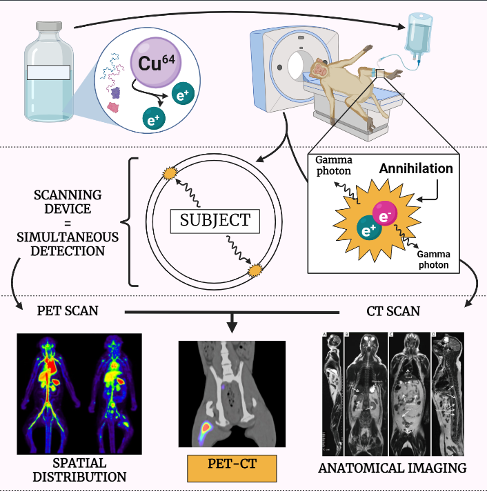
- When a positron is emitted, it travels a short distance and then collides with an electron, resulting in the simultaneous emission of two gamma rays in opposite directions.
- These gamma rays can be detected by a specialized camera called a PET scanner, which uses these gamma rays to create detailed images of the body’s internal structures and biological processes.
Radiotracers in PET (Positron Emission Tomography):
- In order to perform a PET scan, a small amount of a radioactive material, called a radiotracer, is introduced into the body.
- The radiotracer is designed to target specific organs or tissues, and as it travels through the body, it emits positrons which are then detected by the PET scanner.
- The most common radiotracers used in PET imaging are FDG, which is used to detect cancer, and O-15 water, which is used to detect blood flow and metabolism in the brain.
Applications:
- PET imaging is used to detect and diagnose a wide range of diseases and conditions, including cancer, heart disease, and brain disorders.
- It is also used to monitor the effectiveness of treatment, to detect recurrence of cancer and to evaluate the response to therapy.
- It has also been used in research to study the brain and to evaluate the effects of drugs on the brain.
Limitations and Challenges:
- One of the main limitations of PET imaging is that it requires the use of radioactive materials, which can be associated with some risks and safety concerns.
- Additionally, PET imaging is relatively expensive and may not be covered by all insurance plans.
- It also requires specialized equipment and trained personnel, which can limit its availability in certain areas.
- Another limitation is that it has limited spatial resolution compared to other imaging techniques such as MRI.
Data Analysis and Interpretation:
- The data obtained from a PET scan is complex and requires specialized software and trained personnel to analyze and interpret.
- The images are typically presented in color-coded format, with different colors representing different levels of radioactive tracer uptake.
- The images are analyzed for any abnormal uptake of the tracer which could indicate the presence of disease or abnormal biological processes.
Conclusion:
- Positron emission tomography (PET) is a nuclear medicine imaging technique that uses small amounts of radioactive materials to produce images of the body’s biological processes.
- It is a non-invasive diagnostic tool that can be used to detect and diagnose a wide range of diseases and conditions, including cancer, heart disease, and brain disorders.
- Despite its limitations and challenges, PET imaging remains an important diagnostic tool in the field of medicine and is used in both clinical practice and research.
