Table of Contents
Introduction to Phase Contrast Microscopy:
A phase contrast microscopy is a type of microscopy that uses a special technique to enhance the contrast of a sample. It is an essential tool for many fields of science and technology, including biology, chemistry, and materials science.
Definition of Phase Contrast Microscopy:
A phase contrast microscope is a microscope that uses a special optical system to enhance the contrast of a sample. It allows to observe samples that are otherwise difficult to see with a regular microscope.
Discovery of Phase Contrast Microscopy:
- The phase contrast microscope was first developed in the 1930s by Frits Zernike.
- Zernike was awarded the Nobel Prize in Physics in 1953 for his work in developing the phase contrast microscope.
Principal:
- The phase contrast microscope works by using a special optical system to enhance the contrast of a sample.
- The special optical system uses a ring-shaped aperture to adjust the phase of the light that passes through the sample.
- This allows for the observation of samples that are otherwise difficult to see with a regular microscope.
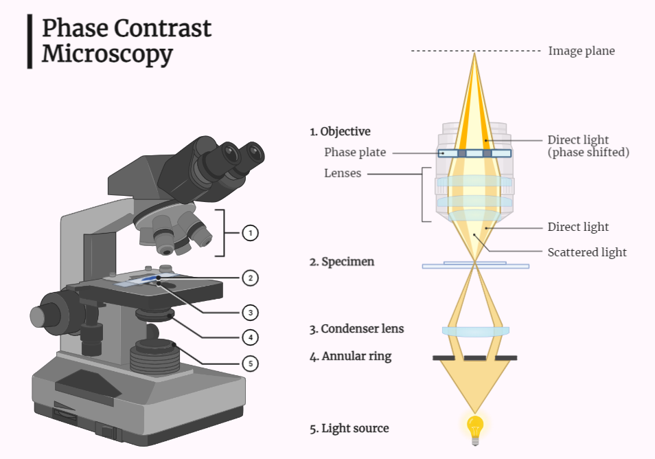
Components:
- The main components of a phase contrast microscope include the light source, the objective lens, the condenser, the phase ring, and the eyepiece lens.
- The light source is used to illuminate the sample.
- The objective lens focuses the light on the sample.
- The condenser focuses the light on the sample and controls the aperture.
- The phase ring is used to adjust the phase of the light that passes through the sample.
- The eyepiece lens magnifies the image formed by the objective lens.
Steps:
- The first step in using a phase contrast microscope is to prepare the sample. This can involve staining the sample or cutting it into thin slices.
- Next, the sample is placed on the microscope stage and the light source is turned on.
- The objective lens is adjusted to focus on the sample and the eyepiece lens is used to magnify the image.
- The phase ring is adjusted to enhance the contrast of the image.
- The image can be viewed through the eyepiece or can be captured by a camera.
Applications:
- Phase contrast microscopes are used in a wide variety of fields, including biology, chemistry, and materials science.
- They are used to study the structure and composition of cells and other biological samples, to analyze chemical compounds, and to study the properties of materials.
- Phase contrast microscopes are indispensable tools in various scientific fields. Their ability to enhance contrast reveals intricate details, driving advancements in biology, chemistry, and materials science.