Table of Contents
Introduction to Transmission Electron Microscope (TEM)
The transmission electron microscope (TEM) is a pioneering technology in electron microscopy, first developed by Knoll and Ruska in the 1930s. This type of microscope allows for the observation of specimens at a molecular level by focusing electrons on a thin sample and detecting those that are transmitted through the specimen.
Working Principle of TEM
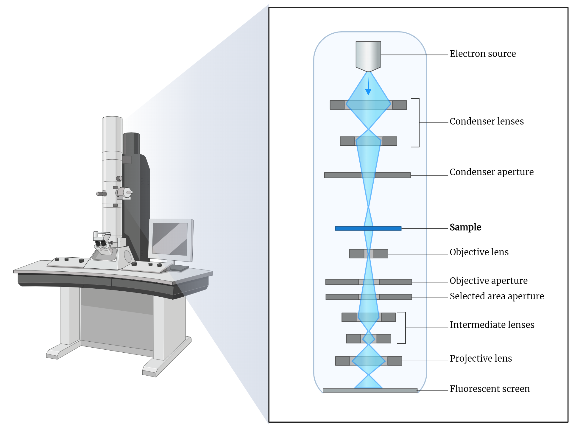
Electron Gun and Acceleration: TEMs typically use thermionic emission guns, which accelerate electrons to potentials ranging from 40 to 200 kV. Advanced TEMs can achieve potentials exceeding 1000 kV, providing higher resolutions. Recently, field emission guns have gained popularity due to their brightness and the finely focused electron beams they produce.
Electron Focusing and Detection: In a TEM, electrons are focused on a thin specimen. As these electrons pass through the specimen, their interaction provides detailed insights into the sample’s structure. The transmitted electrons are detected and used to generate highly detailed images.
Sample Preparation for TEM
Specimen Thinness: One of the key requirements for TEM analysis is that specimens must be extremely thin. Similar to SEM, the samples need to be fixed and dried before analysis. This preparation process can be intricate and labor-intensive.
Fixation and Dehydration: The samples are typically fixed using a combination of glutaraldehyde and paraformaldehyde. Secondary staining is often done with osmium tetroxide (OsO4), which fixes unsaturated lipids and acts as an electron stain due to its heavy metal properties. The specimens are then dehydrated in a manner similar to SEM analysis, using an increasing gradient of ethanol until they are fully dehydrated.
Sectioning and Embedding: The dried samples are then sectioned to obtain ultrathin sections, usually less than 100 nm thick. This is typically achieved by embedding the sample in a plastic mold and cutting it into thin sections. Epoxy and acrylic resins are also used for embedding during sectioning. These ultrathin sections are essential for allowing electrons to pass through the specimen in the TEM.
Staining and Grid Preparation: The sections are stained with heavy metal stains such as uranyl acetate and phosphotungstic acid to enhance contrast. Finally, the stained sample is placed on a carbon-coated grid for TEM analysis.
Scanning Transmission Electron Microscopy (STEM)
Combination of Techniques: STEM combines the principles of SEM and TEM. It operates in scanning mode like an SEM but also allows for the detection of transmitted electrons. An electron beam is focused to a small spot and scanned across the specimen.
Detection Capabilities: STEM is capable of detecting transmitted, secondary, and backscattered electrons, providing spatially resolved information about the specimen. This makes STEM a versatile tool for detailed analysis of specimen structure and composition.
Conclusion
The transmission electron microscope (TEM) is a groundbreaking tool in the field of electron microscopy, offering unparalleled insights into the molecular and atomic structure of specimens. With advancements in electron gun technology and detailed sample preparation techniques, TEM continues to be a crucial instrument in scientific research. The integration of TEM and SEM principles in STEM further enhances the capabilities of electron microscopy, allowing for comprehensive analysis of specimens.