-
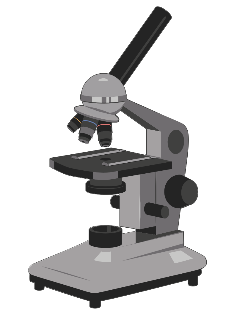
Applications of Light Microscopy
Light microscopy has revolutionized the study of cellular structures and organization within the biological sciences. From simple cell identification and quality assessments to more advanced applications like protein dynamics and co-localization of proteins, this powerful tool plays a crucial role. Techniques such as phase contrast and fluorescence microscopy have significantly enhanced the visualization of specimens,…
-
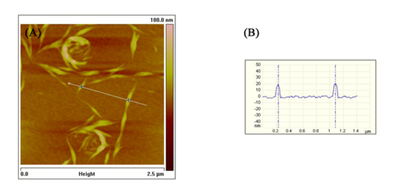
Applications of Atomic Force Microscopy
Atomic Force Microscopy (AFM) is a versatile tool used for detailed surface imaging and analysis. It offers high-resolution imaging for both dry and liquid samples, making it invaluable for biological studies, nucleic acid research, and mechanical property analysis. AFM’s applications range from real-time cell biology observations to nanofabrication and defect detection in materials, highlighting its…
-
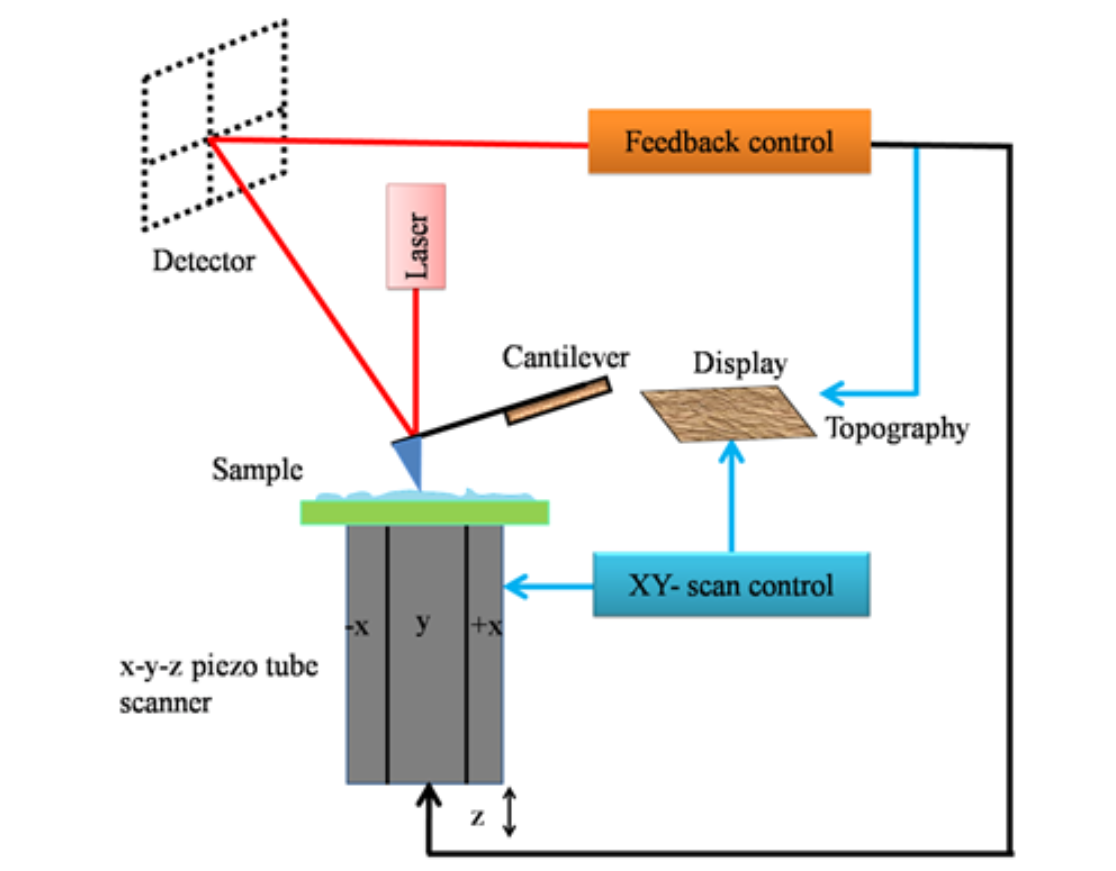
Atomic Force Microscopy
Introduction to Atomic Force Microscopy (AFM) Atomic force microscopy (AFM) is a cutting-edge member of the microscopic techniques known collectively as scanning probe microscopy (SPM). The underlying principles of SPM are markedly distinct from those of light and electron microscopy. SPM methods are used to study the surface properties of materials by scanning an extremely fine probe over the specimen surface. This relatively recent technique emerged in 1981 when Gerd Binnig and Heinrich Rohrer developed the first working SPM, specifically the scanning tunneling microscope (STM). Fundamentals of Scanning Probe Microscopy (SPM) Atomic Force Microscope (AFM) Modes of AFM Operation Imaging Modes Force Mode AFM/Force Spectroscopy Resolution of AFM Advantages of AFM Conclusion Atomic force microscopy (AFM) represents a significant advancement in microscopic techniques, enabling detailed surface analysis and manipulation at the nanoscale. With its versatile operation modes and superior resolution capabilities, AFM stands out as an indispensable tool in scientific research, offering unique advantages over traditional electron microscopy, especially in the study of soft and biological samples.
-
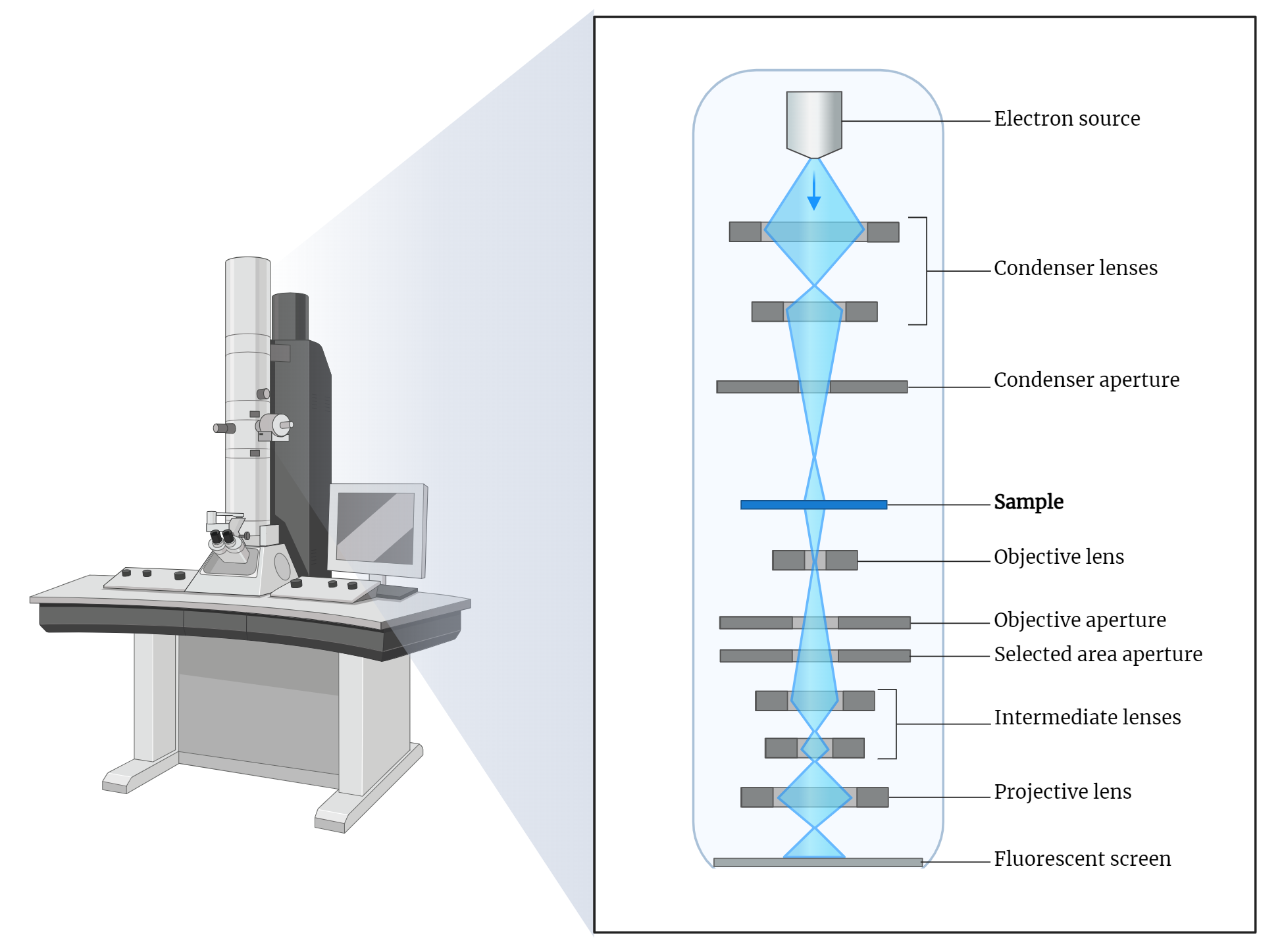
Transmission Electron Microscope (TEM)
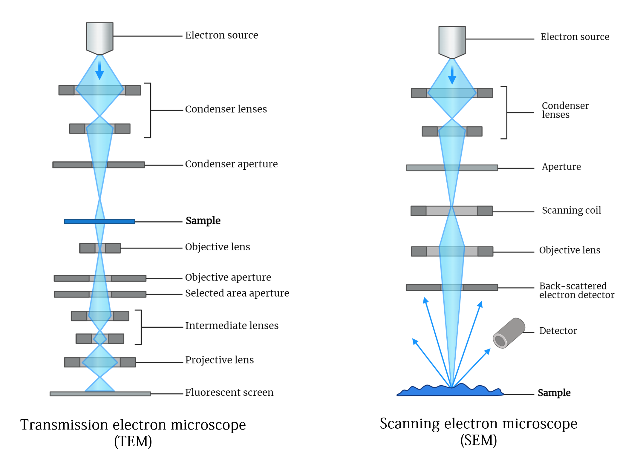
The transmission electron microscope (TEM) is a groundbreaking tool in electron microscopy, first developed by Knoll and Ruska in the 1930s. It utilizes a focused electron beam to produce highly detailed images of thin specimens by detecting electrons transmitted through them. TEMs employ thermionic emission guns and can accelerate electrons to high potentials, enabling high-resolution…
-
Scanning Electron Microscope (SEM)
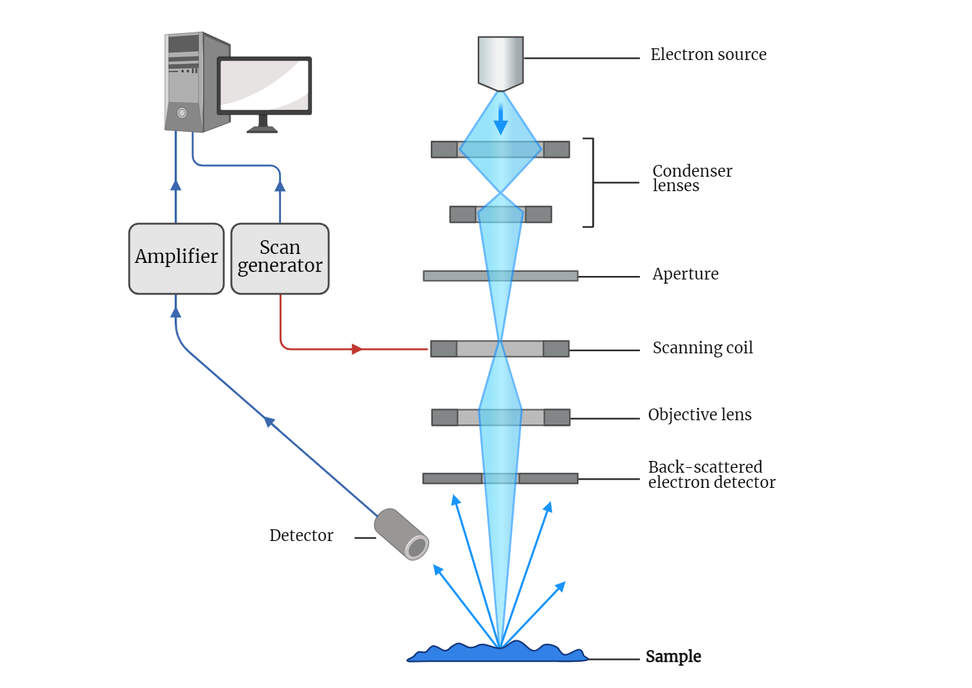
The scanning electron microscope (SEM) is a revolutionary imaging tool that allows for detailed observation of specimen surfaces at a microscopic level. Utilizing a focused beam of electrons, SEMs generate high-resolution images that reveal fine surface structures. The electron gun and magnetic lenses direct and focus the electrons onto the specimen, which is then scanned…
-
Electron Microscopy
In 1924, Louis de Broglie theorized that particles possess wave-like properties, a concept later confirmed by electron diffraction experiments. This breakthrough laid the foundation for electron microscopy. The de Broglie equation describes the relationship between a particle’s wavelength and its momentum, highlighting that electrons accelerated in a microscope can achieve extremely short wavelengths, crucial for…
-
Multiphoton Laser Scanning Microscopy
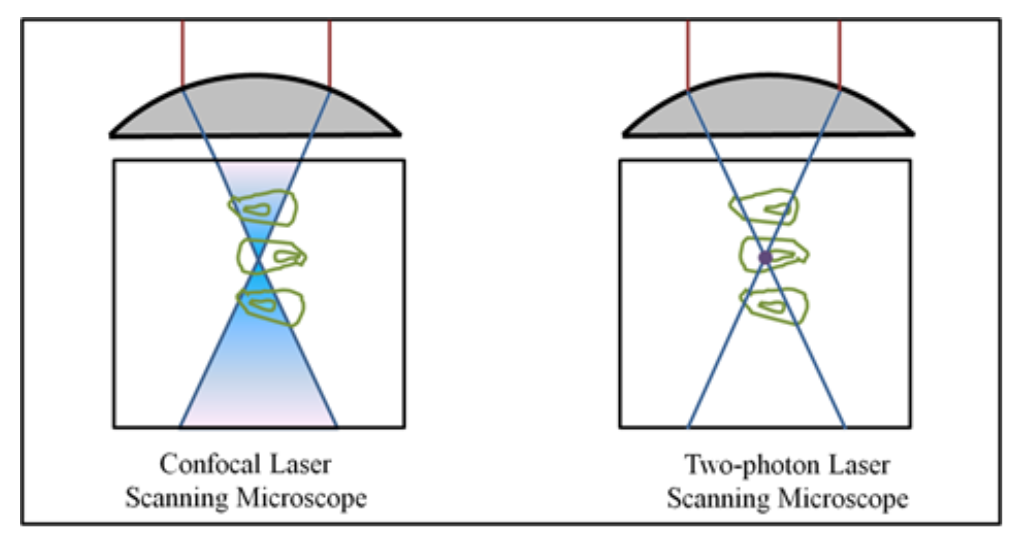
Multiphoton microscopy utilizes the simultaneous absorption of two photons to excite fluorophores, enabling imaging with longer wavelength light. The process necessitates intense light sources, such as pulsed infrared lasers, to achieve sufficient photon density. Notably, this technique allows deeper tissue imaging, as biological specimens poorly absorb near-infrared radiation compared to UV and blue-green radiation. Additionally,…
-
CLSM -Confocal Laser Scanning Microscopy
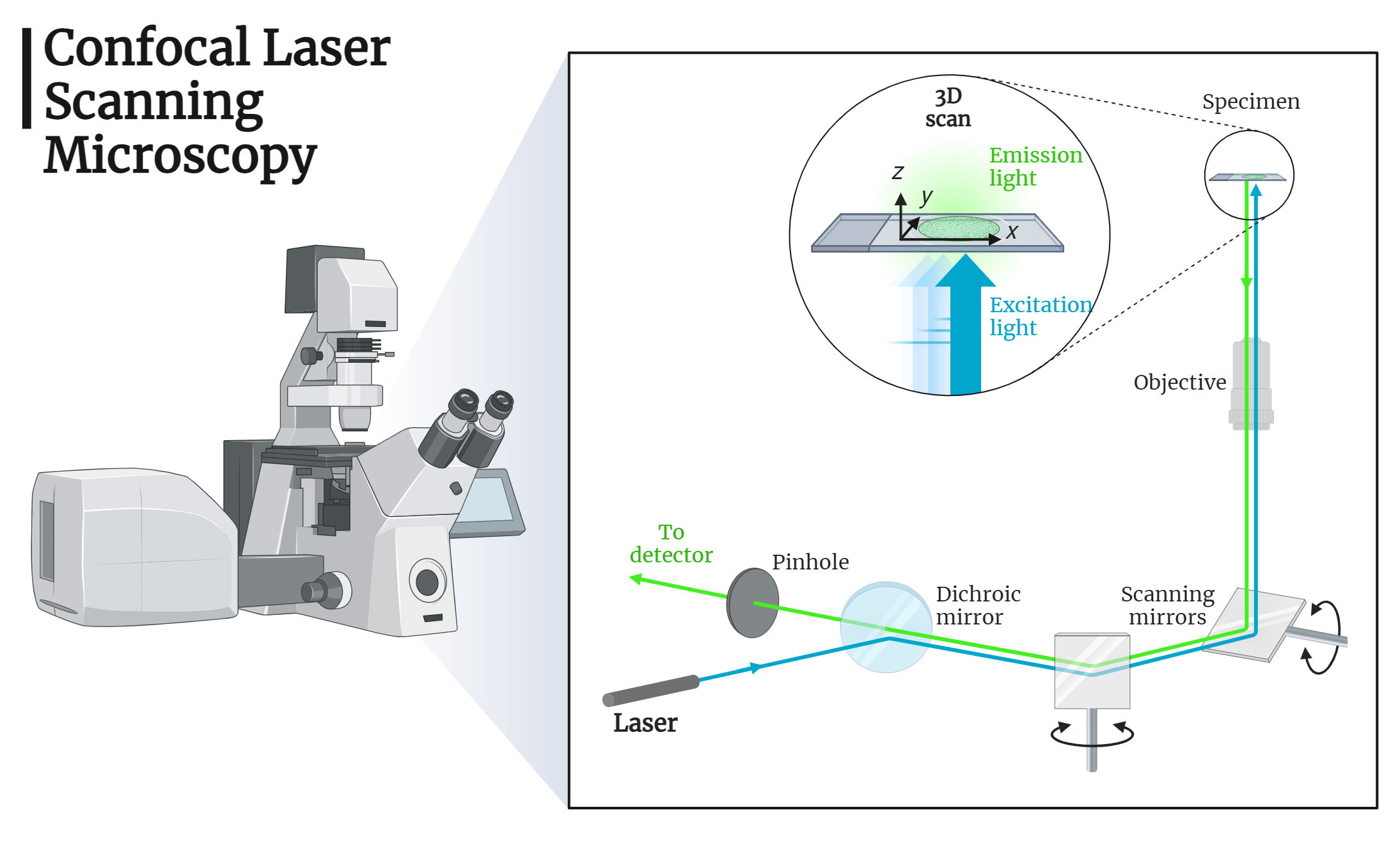
Confocal Laser Scanning Microscopy (CLSM) enables high-resolution imaging of biological specimens by focusing on thin layers within the sample. Utilizing lasers, pinhole apertures, and raster scanning, CLSM provides sharp, detailed images and facilitates three-dimensional reconstruction. This technique offers significant advantages in the precise localization and visualization of cellular structures, paving the way for advancements in…
-
Fluorescence Microscopy
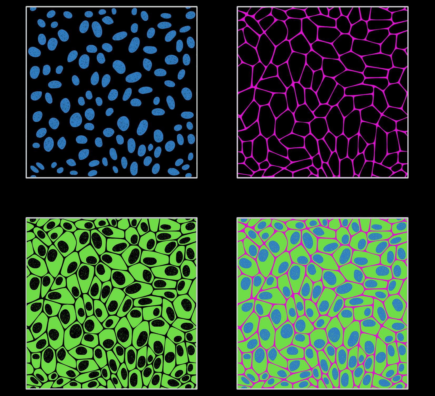
Fluorescence microscopy is a pivotal technique in biological research, allowing the visualization of cellular structures and functions with enhanced contrast and resolution. By using fluorescent probes, GFP tagging, and advanced methods like TIRF, researchers can gain detailed insights into cellular processes. This study note covers the principles, types, and applications of fluorescence microscopy, highlighting its…
-
Isolation and Purification of RNA
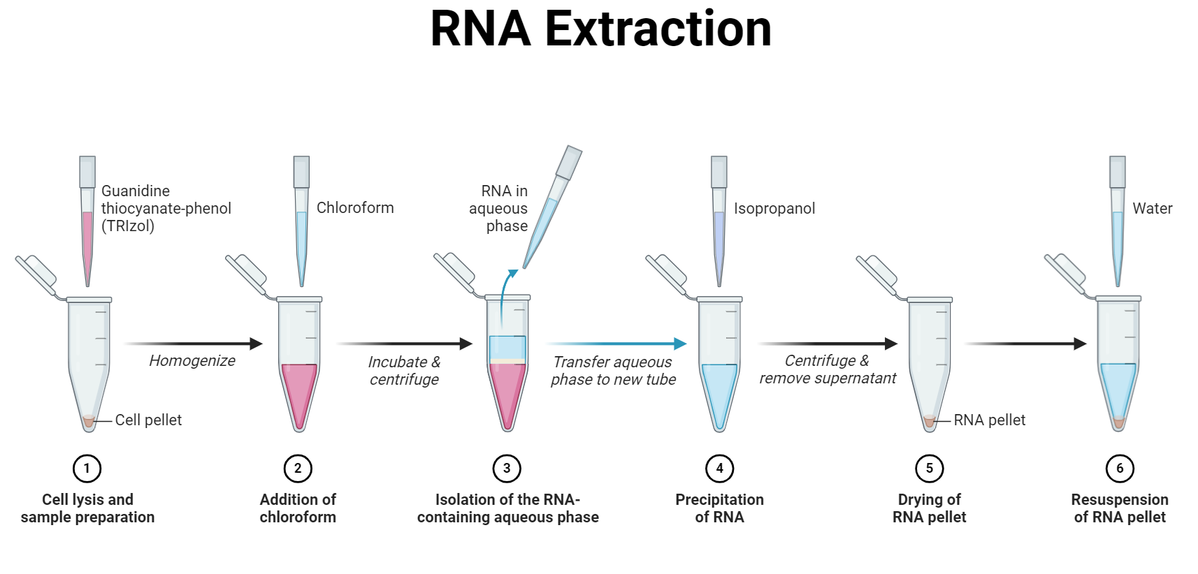
Pure RNA isolation is essential in molecular biology for gene expression studies and downstream analyses. There are two main strategies: total RNA isolation and mRNA isolation, with total RNA isolation being less laborious. Methods include phenol-chloroform extraction, spin column purification, and magnetic bead-based methods, each having unique advantages. Choose based on sample specifics.
-
Spectroscopy and Beer-Lambert’s Law
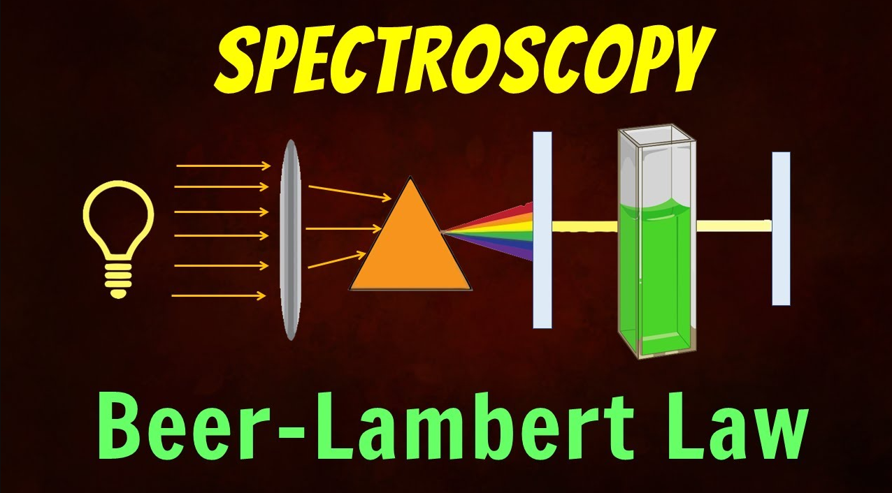
Spectroscopy, a fascinating scientific technique, delves into the captivating interaction between matter and electromagnetic radiation. By analyzing how substances interact with light at different wavelengths, spectroscopy provides crucial insights into the composition, structure, and behavior of materials. One of the fundamental principles in spectroscopy is the Beer-Lambert’s Law, a cornerstone in quantitative analysis. This law…
-

Ion Exchange Chromatography
Introduction Ion exchange chromatography (IEC) is a type of liquid chromatography that separates molecules based on their charge. It is a widely used technique in biochemistry and analytical chemistry for the purification and isolation of biomolecules such as proteins, nucleic acids, and small molecules. Principles of Ion Exchange Chromatography IEC utilizes a stationary phase, which…
-

Nuclear Magnetic Resonance (NMR)
The Nuclear Magnetic Resonance (NMR) technique is a powerful tool used in biophysics to study the structure, dynamics, and interactions of biomolecules in solution. NMR spectroscopy is based on the principle that certain atoms’ nuclei can absorb and emit electromagnetic radiation in the radiofrequency range when placed in a strong magnetic field. This technique can…
-
X-ray Diffraction Technique
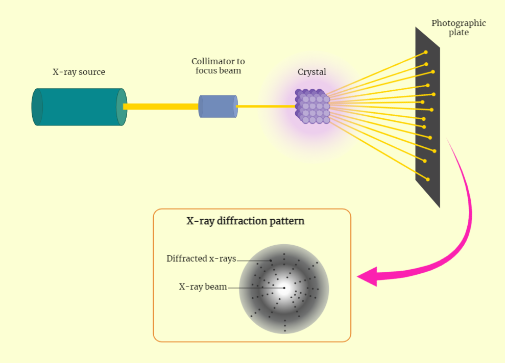
The X-ray diffraction (XRD) technique is a powerful tool for studying the structure of crystalline materials. It works by shining X-rays on a crystalline material and detecting the diffracted X-rays on a detector, creating a pattern of diffraction peaks. The angle and intensity of these peaks can be used to determine the crystal structure, unit…
-
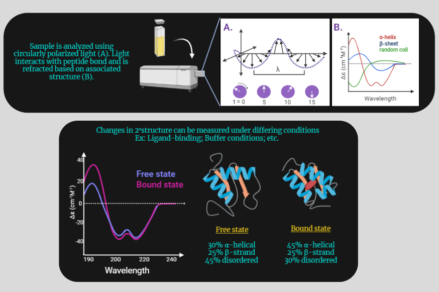
Circular Dichroism (CD) Technique
Circular dichroism (CD) is a powerful spectroscopic technique used to study the structure and properties of chiral molecules, such as proteins, nucleic acids, and carbohydrates. It is based on the differential absorption of left and right circularly polarized light by chiral molecules, which can provide information on their secondary, tertiary, and quaternary structures. In this…
-
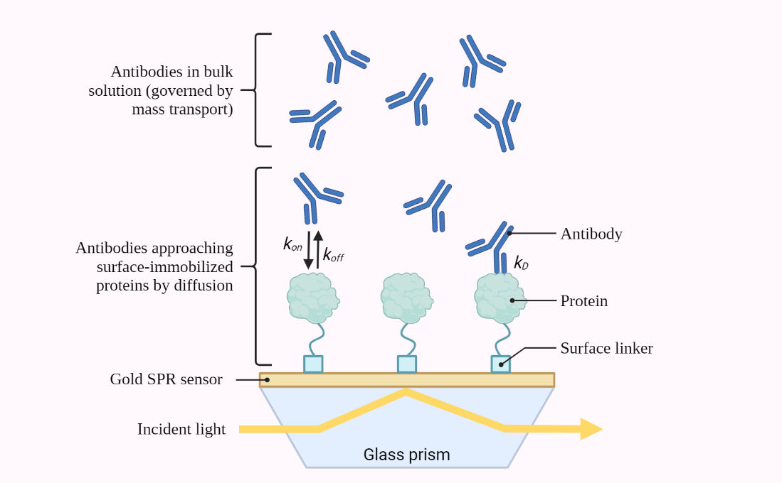
SURFACE PLASMON RESONANCE (SPR)
Surface Plasmon Resonance (SPR) is a powerful label-free technique used for real-time monitoring of biomolecular interactions. This article provides a comprehensive study of the SPR technique, covering its principles, instrumentation, applications, factors affecting the SPR response, advantages and limitations, future directions, and more. With 15 headings and FAQs, this article is a valuable resource for…
-
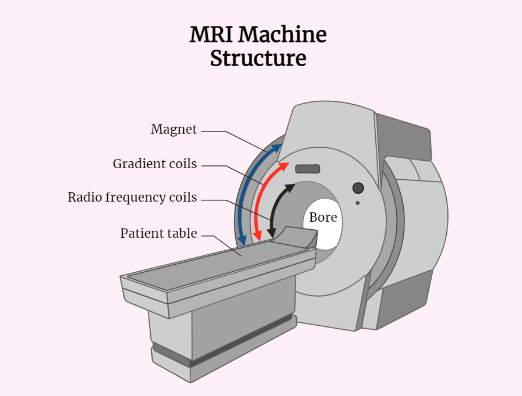
Magnetic Resonance Imaging (MRI)
Magnetic Resonance Imaging (MRI) is a medical imaging technique that creates detailed images of the internal structures of the body using magnetic fields and radio waves. MRI is a non-invasive diagnostic tool that provides high-quality images of soft tissues, organs, and bones without exposing patients to harmful radiation. The basic principle of MRI is to…
-
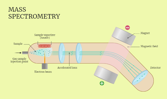
Mass Spectrometry (MS) Technique
The article discusses the principles, techniques, and applications of Mass Spectrometry (MS) from a biophysics perspective. It explains how MS is used to study the structure, dynamics, and interactions of biomolecules and how it can be applied to a wide range of biomolecules, including proteins, nucleic acids, lipids, and small molecules. The article also discusses…
-
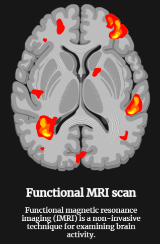
Functional Magnetic Resonance Imaging
Functional Magnetic Resonance Imaging (fMRI) is a powerful medical imaging technique used to study brain function. It measures changes in blood flow in the brain using MRI technology, which can be used to infer changes in neural activity. fMRI is commonly used in neuroscience, psychology, and psychiatry to investigate a wide range of questions related…
-
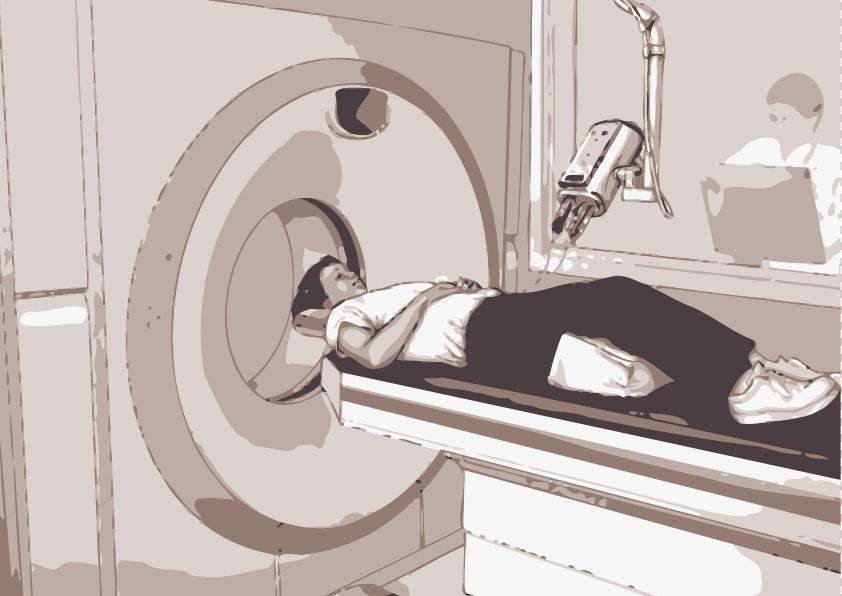
CT Scan Technique
CT (Computed Tomography) scan is a widely used medical imaging technique that produces detailed, cross-sectional images of the body using x-rays and computer technology. The technique uses x-ray detectors, reconstruction algorithms and dose management techniques to minimize the radiation dose to the patient while producing high-quality images. Specialized CT scans such as CT angiography, CT…
-
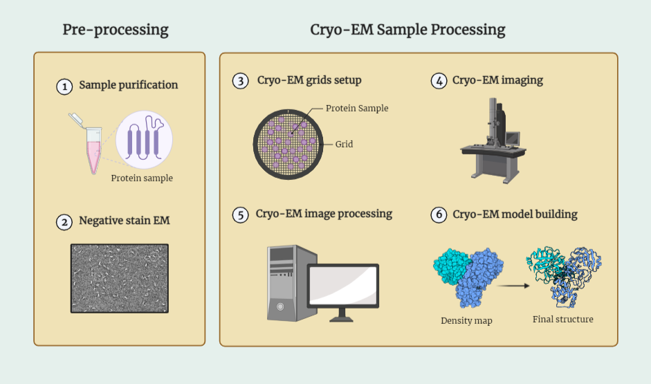
Cryo Electron Microscopy (Cryo-EM)
Cryo-EM is a cutting-edge imaging technique used for studying the structure of biological macromolecules such as proteins and nucleic acids. By using a cryogenic electron microscope, researchers can capture high-resolution images of frozen, hydrated samples, preserving their native state and avoiding radiation damage. The sample preparation is a crucial step that involves rapid freezing of…
-
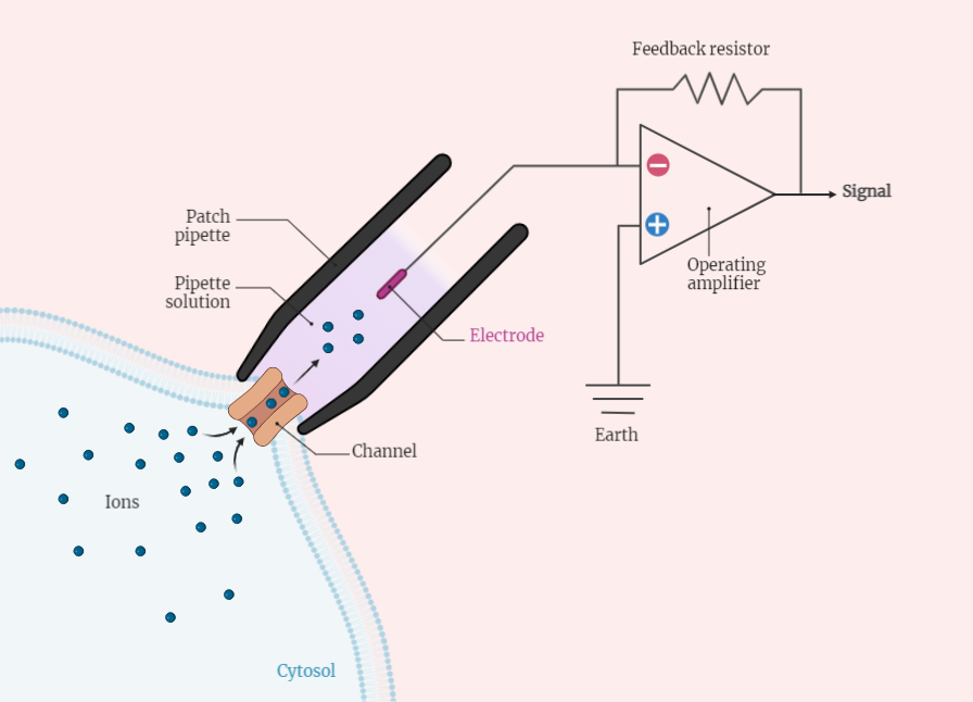
Patch Clamp Technique
The patch clamp technique is a widely used electrophysiological method for studying ion channels in cell membranes. This technique involves using a micropipette to form a gigaohm seal with a cell membrane, allowing for the manipulation of the electrical potential across the membrane. There are four main types of patch clamp, each with their own…
-
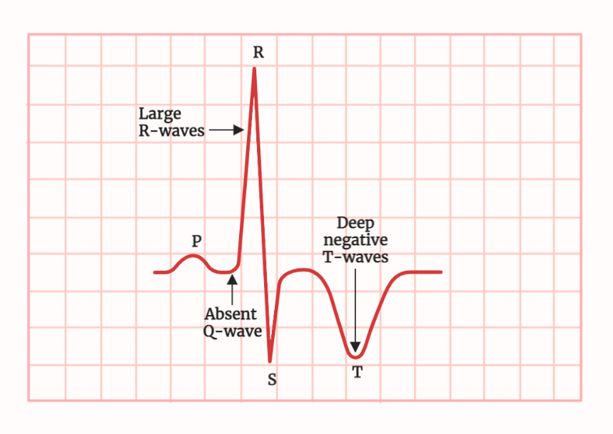
ECG (Electrocardiogram)
The electrocardiogram (ECG) is a widely used diagnostic tool for the evaluation of heart conditions. It is a non-invasive test that records the electrical activity of the heart and displays it as waves on a graph. The ECG is used to diagnose a variety of heart conditions such as arrhythmias, heart attacks, and other cardiac…
-
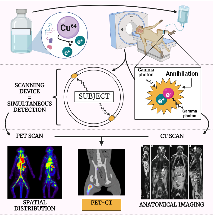
PET (Positron Emission Tomography)
The article provides an in-depth study note on Positron Emission Tomography (PET), a non-invasive nuclear medicine imaging technique that uses small amounts of radioactive materials to produce images of the body’s biological processes. It explains the principles of the PET imaging technique, the use of radiotracers, its applications in detecting and diagnosing various diseases, and…
-

Beer-Lambert Law and Spectrophotometry
Discover the principles of the Beer-Lambert Law and its application in spectrophotometry. Explore how this law relates the concentration of a solute to absorbance, and its significance in quantitative analysis, chemical kinetics, environmental monitoring, pharmaceutical analysis, and biochemical assays. Unlock the power of spectrophotometry in understanding chemical substances and their characteristics.
Categories
- Anatomy (9)
- Animal Form and Functions (38)
- Animal Physiology (65)
- Biochemistry (33)
- Biophysics (25)
- Biotechnology (52)
- Botany (42)
- Plant morphology (6)
- Plant Physiology (26)
- Cell Biology (107)
- Cell Cycle (14)
- Cell Signaling (21)
- Chemistry (9)
- Developmental Biology (36)
- Fertilization (13)
- Ecology (5)
- Embryology (17)
- Endocrinology (10)
- Environmental biology (3)
- Genetics (59)
- DNA (27)
- Inheritance (13)
- Histology (3)
- Hormone (3)
- Immunology (29)
- life science (76)
- Material science (8)
- Microbiology (18)
- Virus (8)
- Microscopy (18)
- Molecular Biology (113)
- parasitology (6)
- Physics (3)
- Physiology (11)
- Plant biology (26)
- Uncategorized (7)
- Zoology (112)
- Classification (6)
- Invertebrate (7)


