-
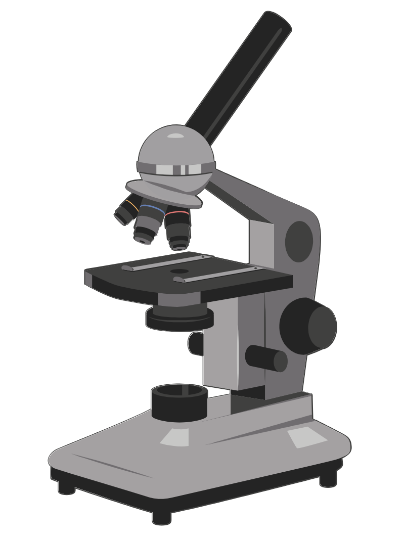
Applications of Light Microscopy
Light microscopy has revolutionized the study of cellular structures and organization within the biological sciences. From simple cell identification and quality assessments to more advanced applications like protein dynamics and co-localization of proteins, this powerful tool plays a crucial role. Techniques such as phase contrast and fluorescence microscopy have significantly enhanced the visualization of specimens,…
-
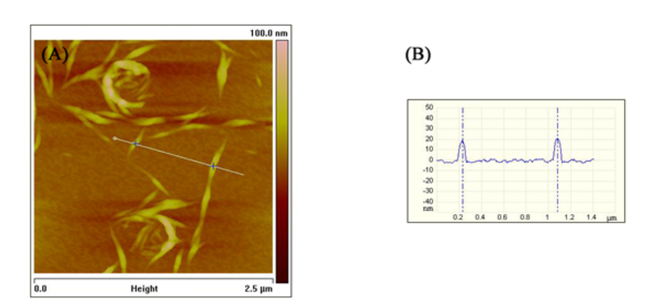
Applications of Atomic Force Microscopy
Atomic Force Microscopy (AFM) is a versatile tool used for detailed surface imaging and analysis. It offers high-resolution imaging for both dry and liquid samples, making it invaluable for biological studies, nucleic acid research, and mechanical property analysis. AFM’s applications range from real-time cell biology observations to nanofabrication and defect detection in materials, highlighting its…
-
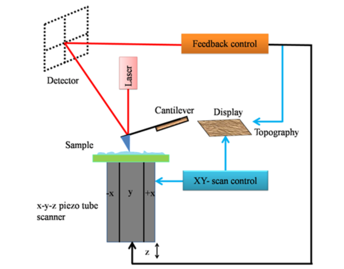
Atomic Force Microscopy
Introduction to Atomic Force Microscopy (AFM) Atomic force microscopy (AFM) is a cutting-edge member of the microscopic techniques known collectively as scanning probe microscopy (SPM). The underlying principles of SPM are markedly distinct from those of light and electron microscopy. SPM methods are used to study the surface properties of materials by scanning an extremely fine probe over the specimen surface. This relatively recent technique emerged in 1981 when Gerd Binnig and Heinrich Rohrer developed the first working SPM, specifically the scanning tunneling microscope (STM). Fundamentals of Scanning Probe Microscopy (SPM) Atomic Force Microscope (AFM) Modes of AFM Operation Imaging Modes Force Mode AFM/Force Spectroscopy Resolution of AFM Advantages of AFM Conclusion Atomic force microscopy (AFM) represents a significant advancement in microscopic techniques, enabling detailed surface analysis and manipulation at the nanoscale. With its versatile operation modes and superior resolution capabilities, AFM stands out as an indispensable tool in scientific research, offering unique advantages over traditional electron microscopy, especially in the study of soft and biological samples.
-
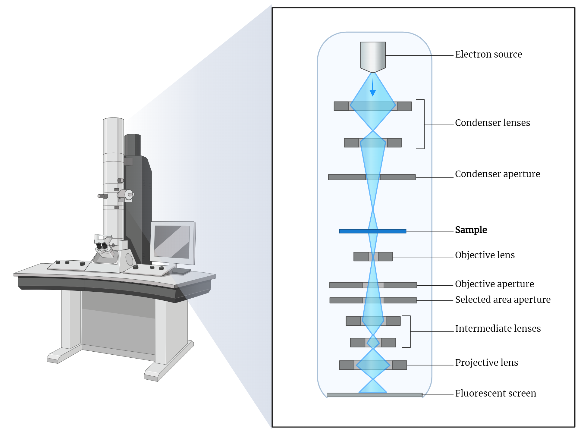
Transmission Electron Microscope (TEM)
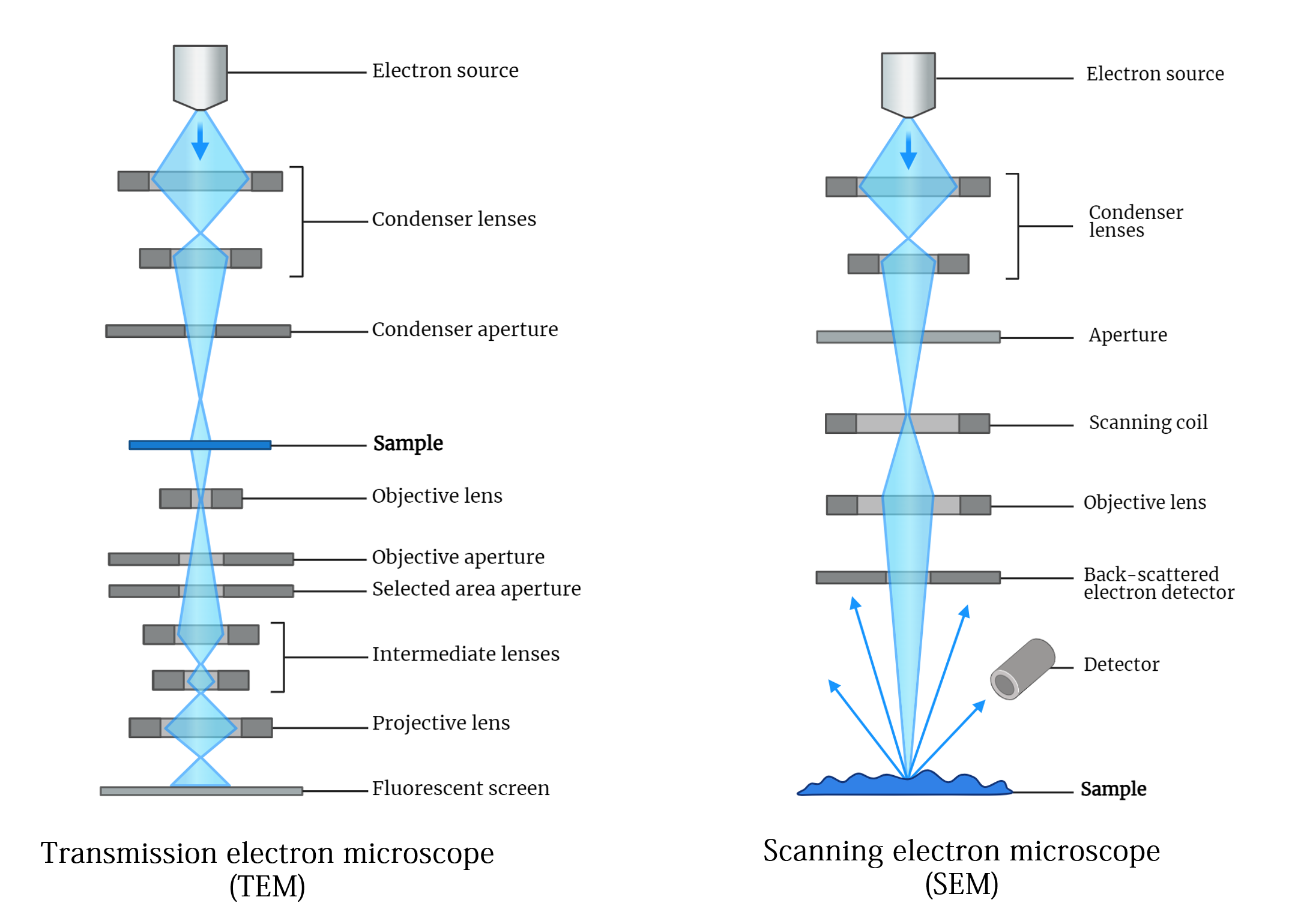
The transmission electron microscope (TEM) is a groundbreaking tool in electron microscopy, first developed by Knoll and Ruska in the 1930s. It utilizes a focused electron beam to produce highly detailed images of thin specimens by detecting electrons transmitted through them. TEMs employ thermionic emission guns and can accelerate electrons to high potentials, enabling high-resolution…
-
Scanning Electron Microscope (SEM)
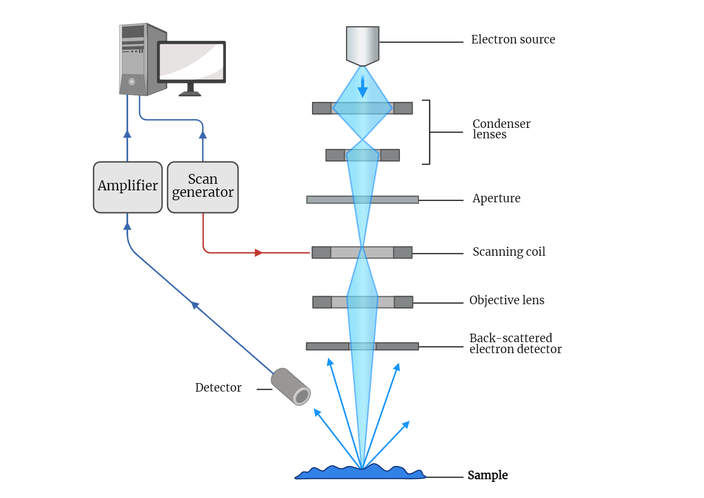
The scanning electron microscope (SEM) is a revolutionary imaging tool that allows for detailed observation of specimen surfaces at a microscopic level. Utilizing a focused beam of electrons, SEMs generate high-resolution images that reveal fine surface structures. The electron gun and magnetic lenses direct and focus the electrons onto the specimen, which is then scanned…
-
Electron Microscopy
In 1924, Louis de Broglie theorized that particles possess wave-like properties, a concept later confirmed by electron diffraction experiments. This breakthrough laid the foundation for electron microscopy. The de Broglie equation describes the relationship between a particle’s wavelength and its momentum, highlighting that electrons accelerated in a microscope can achieve extremely short wavelengths, crucial for…
-
Multiphoton Laser Scanning Microscopy
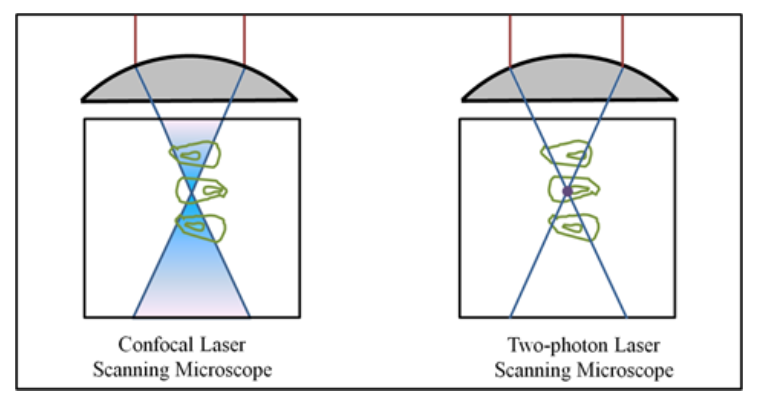
Multiphoton microscopy utilizes the simultaneous absorption of two photons to excite fluorophores, enabling imaging with longer wavelength light. The process necessitates intense light sources, such as pulsed infrared lasers, to achieve sufficient photon density. Notably, this technique allows deeper tissue imaging, as biological specimens poorly absorb near-infrared radiation compared to UV and blue-green radiation. Additionally,…
-
CLSM -Confocal Laser Scanning Microscopy
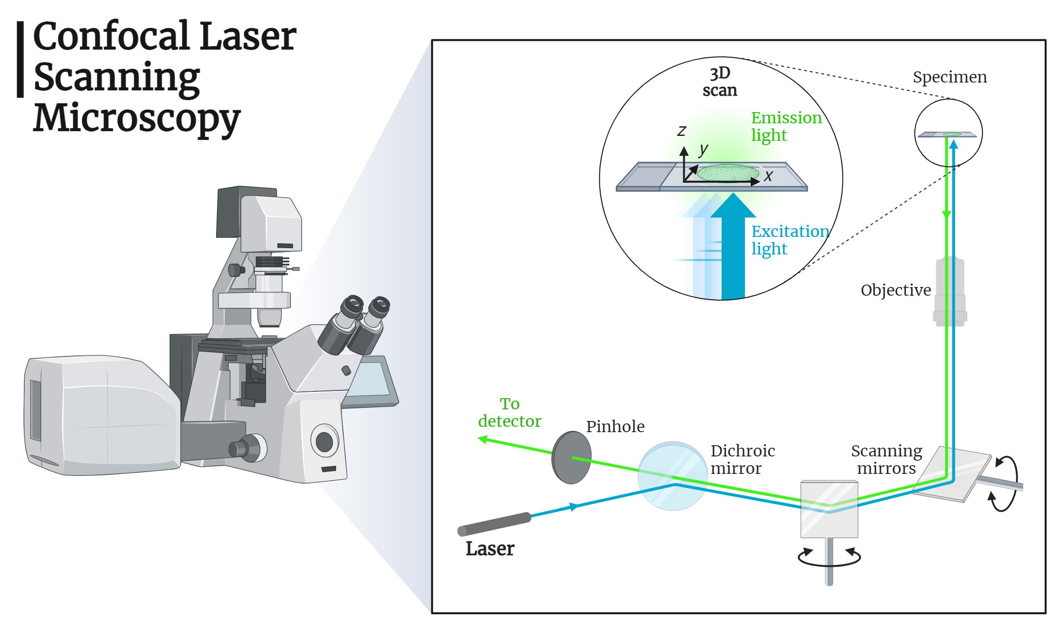
Confocal Laser Scanning Microscopy (CLSM) enables high-resolution imaging of biological specimens by focusing on thin layers within the sample. Utilizing lasers, pinhole apertures, and raster scanning, CLSM provides sharp, detailed images and facilitates three-dimensional reconstruction. This technique offers significant advantages in the precise localization and visualization of cellular structures, paving the way for advancements in…
-
Fluorescence Microscopy
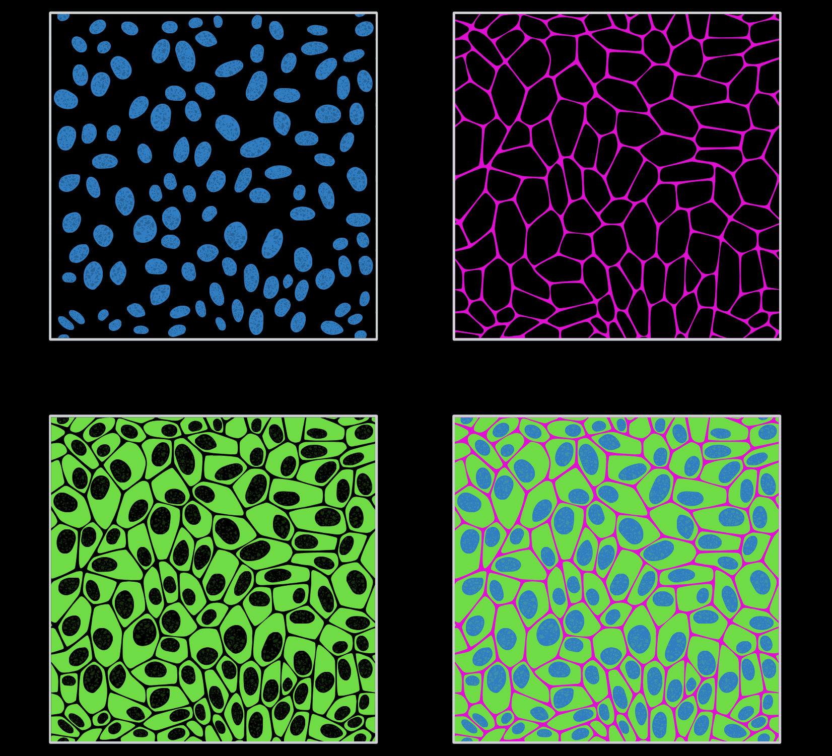
Fluorescence microscopy is a pivotal technique in biological research, allowing the visualization of cellular structures and functions with enhanced contrast and resolution. By using fluorescent probes, GFP tagging, and advanced methods like TIRF, researchers can gain detailed insights into cellular processes. This study note covers the principles, types, and applications of fluorescence microscopy, highlighting its…
-
Microscopy
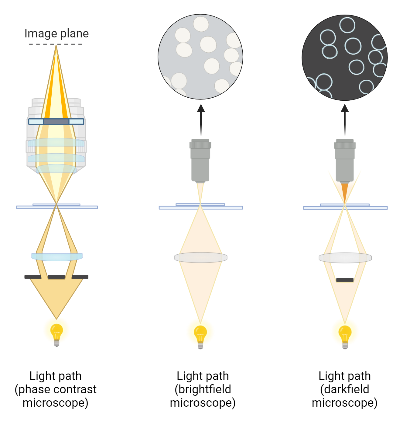
Light microscopy plays a crucial role in biological research, offering a means to visualize microscopic objects and macromolecules. By leveraging the principles of geometrical optics, various forms of microscopy, such as bright-field, dark-field, and phase contrast, enhance image contrast and resolution in unique ways. This study note delves into the fundamentals of light microscopy, including…
-
Objective Lenses of Microscopes
Discover the various types of objective lenses used in microscopes. Learn about achromatic, plan, apochromatic, and oil immersion lenses, their unique properties, and applications in microscopy. Choose the right lens for enhanced imaging and accurate observations.
-
Atomic Force Microscopy (AFM)
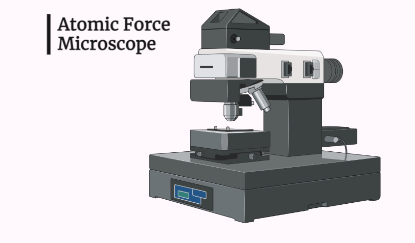
Atomic Force Microscopy (AFM) is a powerful imaging technique used to visualize and manipulate materials at the nanoscale. By scanning a sharp probe over a sample surface, AFM measures the interaction forces between the probe and the sample, providing high-resolution images and information about surface properties. This study note delves into the principles, instrumentation, operational…
-
Phase Contrast Microscopy
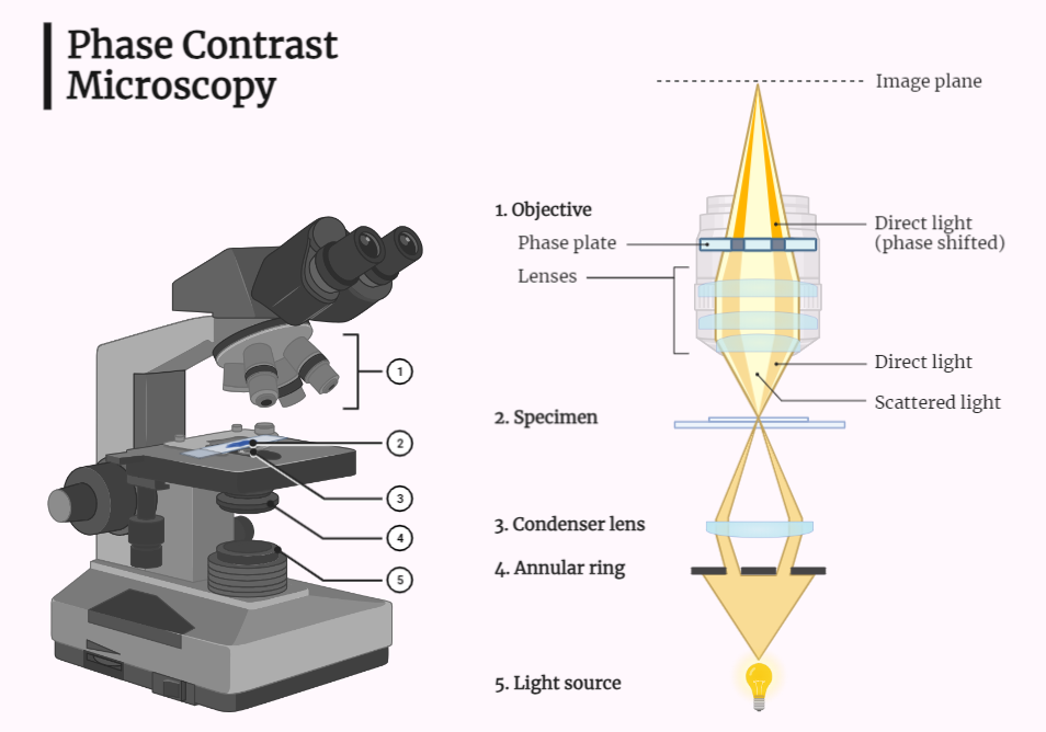
Discover the power of a phase contrast microscope, an essential tool for enhancing sample contrast in fields like biology and materials science. Learn about its components, working principle, and applications in studying cell structures and analyzing materials. Explore the advancements in microscopy techniques and scientific instruments for improved research and imaging capabilities.
-
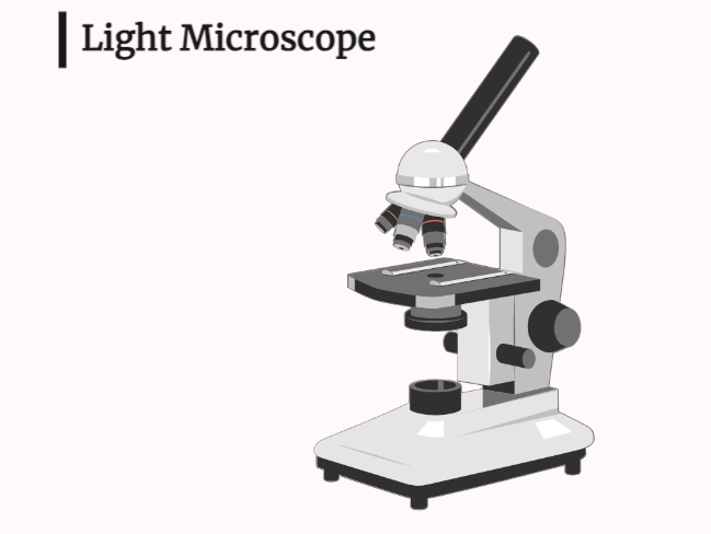
Light Microscopy
Light microscopy is a versatile technique used in various scientific disciplines such as biology, chemistry, and materials science. By illuminating a sample with light and capturing the transmitted image, this essential tool enables the study of cell structure, materials analysis, and scientific research. With its simplicity and ability to produce high-resolution images, light microscopy plays…
-
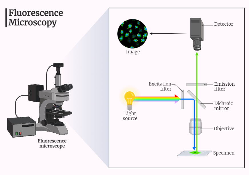
Fluorescence Microscope
A fluorescence microscope is an essential tool in science and technology, enabling high-resolution imaging of samples using fluorescence techniques. It is widely used in biology, chemistry, and materials science for analyzing cell structures, molecular imaging, and sample analysis. By exciting specific molecules and selectively detecting fluorescence, it provides valuable insights into biological samples and their…
-
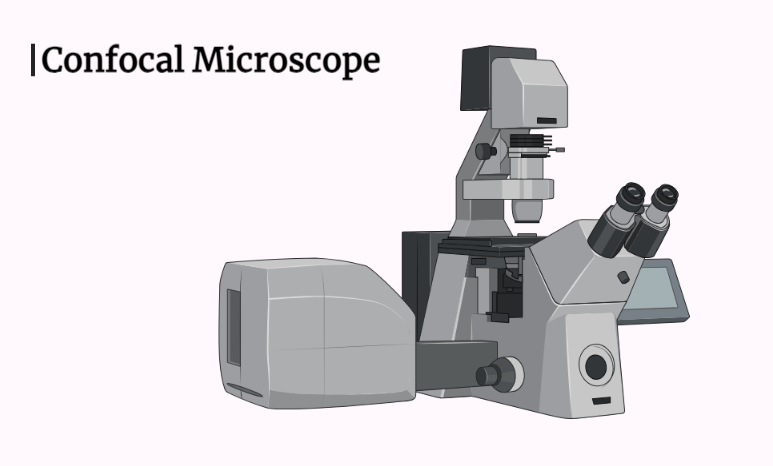
Confocal Microscope
Embark on a fascinating journey into the world of confocal microscopy. Discover its role in high-resolution imaging, sample preparation, and applications in biology, chemistry, and materials science. Uncover the groundbreaking advancements made possible by selectively detecting fluorescence, enabling detailed study of biological samples and components. Witness the transformative power of confocal microscopy in unlocking the…
-
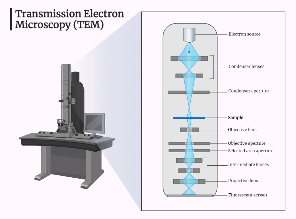
Transmission Electron Microscope (TEM)
Embark on a journey through the world of transmission electron microscopy (TEM). Explore its significance in materials science, biology, and chemistry. Discover the remarkable high-resolution imaging capabilities, sample preparation, and applications across various fields. Uncover the instrumental role of TEM in advancing scientific knowledge and understanding through its ability to reveal the internal structure of…
-

Scanning Electron Microscope (SEM)
Discover the powerful world of scanning electron microscopy (SEM). Explore its working principle, from the electron gun to image analysis. Uncover its applications in materials science, biology, geology, and semiconductor manufacturing. Gain insights into the remarkable high-resolution imaging capabilities of SEM and its significant impact on scientific discoveries and advancements.
Categories
- Anatomy (9)
- Animal Form and Functions (38)
- Animal Physiology (65)
- Biochemistry (33)
- Biophysics (25)
- Biotechnology (52)
- Botany (42)
- Plant morphology (6)
- Plant Physiology (26)
- Cell Biology (107)
- Cell Cycle (14)
- Cell Signaling (21)
- Chemistry (9)
- Developmental Biology (36)
- Fertilization (13)
- Ecology (5)
- Embryology (17)
- Endocrinology (10)
- Environmental biology (3)
- Genetics (59)
- DNA (27)
- Inheritance (13)
- Histology (3)
- Hormone (3)
- Immunology (29)
- life science (76)
- Material science (8)
- Microbiology (18)
- Virus (8)
- Microscopy (18)
- Molecular Biology (113)
- parasitology (6)
- Physics (3)
- Physiology (11)
- Plant biology (26)
- Uncategorized (7)
- Zoology (112)
- Classification (6)
- Invertebrate (7)


