-

Atomic Force Microscopy
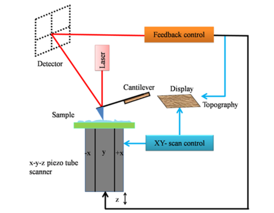
Introduction to Atomic Force Microscopy (AFM) Atomic force microscopy (AFM) is a cutting-edge member of the microscopic techniques known collectively as scanning probe microscopy (SPM). The underlying principles of SPM are markedly distinct from those of light and electron microscopy. SPM methods are used to study the surface properties of materials by scanning an extremely fine probe over the specimen surface. This relatively recent technique emerged in 1981 when Gerd Binnig and Heinrich Rohrer developed the first working SPM, specifically the scanning tunneling microscope (STM). Fundamentals of Scanning Probe Microscopy (SPM) Atomic Force Microscope (AFM) Modes of AFM Operation Imaging Modes Force Mode AFM/Force Spectroscopy Resolution of AFM Advantages of AFM Conclusion Atomic force microscopy (AFM) represents a significant advancement in microscopic techniques, enabling detailed surface analysis and manipulation at the nanoscale. With its versatile operation modes and superior resolution capabilities, AFM stands out as an indispensable tool in scientific research, offering unique advantages over traditional electron microscopy, especially in the study of soft and biological samples.
Categories
- Anatomy (9)
- Animal Form and Functions (38)
- Animal Physiology (65)
- Biochemistry (33)
- Biophysics (25)
- Biotechnology (52)
- Botany (42)
- Plant morphology (6)
- Plant Physiology (26)
- Cell Biology (107)
- Cell Cycle (14)
- Cell Signaling (21)
- Chemistry (9)
- Developmental Biology (36)
- Fertilization (13)
- Ecology (5)
- Embryology (17)
- Endocrinology (10)
- Environmental biology (3)
- Genetics (59)
- DNA (27)
- Inheritance (13)
- Histology (3)
- Hormone (3)
- Immunology (29)
- life science (76)
- Material science (8)
- Microbiology (18)
- Virus (8)
- Microscopy (18)
- Molecular Biology (113)
- parasitology (6)
- Physics (3)
- Physiology (11)
- Plant biology (26)
- Uncategorized (7)
- Zoology (112)
- Classification (6)
- Invertebrate (7)


